. 2010 Feb;84(3):1376-86.
doi: 10.1128/JVI.01220-09. Epub 2009 Nov 18.

Human papillomavirus type 8 E2 protein unravels JunB/Fra-1 as an activator of the beta4-integrin gene in human keratinocytes

Affiliations

Human papillomavirus type 8 E2 protein unravels JunB/Fra-1 as an activator of the beta4-integrin gene in human keratinocytes

Monika Oldak et al. J Virol. 2010 Feb.

Abstract

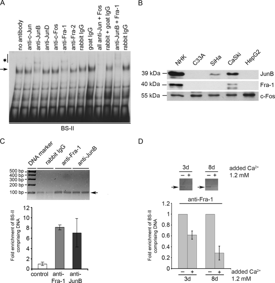
The papillomavirus life cycle parallels keratinocyte differentiation in stratifying epithelia. We have previously shown that the human papillomavirus type 8 (HPV8) E2 protein downregulates beta4-integrin expression in normal human keratinocytes, which may trigger subsequent differentiation steps. Here, we demonstrate that the DNA binding domain of HPV8 E2 is sufficient to displace a cellular factor from the beta4-integrin promoter. We identified the E2-displaceable factor as activator protein 1 (AP-1), a heteromeric transcription factor with differentiation-specific expression in the epithelium. beta4-Integrin-positive epithelial cells displayed strong AP-1 binding activity. Both AP-1 binding activity and beta4-integrin expression were coregulated during keratinocyte differentiation suggesting the involvement of AP-1 in beta4-integrin expression. In normal human keratinocytes the AP-1 complex was composed of JunB and Fra-1 subunits. Chromatin immunoprecipitation assays confirmed that JunB/Fra-1 proteins interact in vivo with the beta4-integrin promoter and that JunB/Fra-1 promoter occupancy is reduced during keratinocyte differentiation as well as in HPV8 E2 positive keratinocytes. Ectopic expression of the tethered JunB/Fra-1 heterodimer in normal human keratinocytes activated the beta4-integrin promoter, while coexpression of HPV8 E2 reverted the JunB/Fra-1 effect. In summary, we identified a novel mechanism of human beta4-integrin regulation that is specifically targeted by the HPV8 E2 protein mimicking transcriptional conditions of differentiation. This may explain the early steps of how HPV8 commits its host cells to the differentiation process required for the viral life cycle.


Figures

FIG. 1.
FIG. 1.
The carboxy terminus of HPV8 E2 displaces a cellular factor binding to the β4-integrin promoter. Nuclear extracts isolated from NHK (NE-NHK) were incubated with 32P-labeled BS-II oligonucleotides and His-tagged HPV8-E2C (left panel) or nuclear extracts containing EYFP-8E2C fusion protein (right panel) and analyzed by EMSA. The E2C specific bands are indicated by stars. The arrow indicates the E2-displaceable cellular factor E2-DF binding to the BS-II oligonucleotides.
FIG. 2.
FIG. 2.
Calcium-induced reduction of β4-integrin expression and E2-DF DNA binding activity. NHK treated with 1.2 and 2.4 mM CaCl2 were harvested after 48 h and subjected to β4-integrin staining and nuclear extract isolation. (A) Expression levels of β4-integrin were determined by flow cytometry. The mean fluorescence intensity of β4-integrin expression in untreated cells was set to 1, and the average fold change from two independent experiments is shown. (B) In EMSA, 32P-labeled BS-II oligonucleotides were incubated with 10 μg of nuclear extracts prepared from NHK cultured under different calcium conditions. The arrow indicates complex(es) corresponding to cellular E2-DF.
FIG. 3.
FIG. 3.
E2-displaceable factor recognizes a nonclassical AP-1 binding site within the β4-integrin promoter. BS-II oligonucleotides were truncated at the 3′ and/or 5′ ends and tested by EMSA for binding of E2-DF. 32P-labeled BS-II (lane 2) and BS-II truncated by 5 bp at the 3′ end (lane 1) and by 5 bp (lane 3), 10 bp (lane 4), 15 bp (lane 5), 20 bp (lane 6), and 25 bp (lane 7) at the 5′ end were incubated with 10 μg of nuclear extracts prepared from NHK (NE-NHK). The arrow indicates protein complexes corresponding to cellular E2-DF-binding activity within BS-II.
FIG. 4.
FIG. 4.
BS-II and AP-1 consensus binding site compete for the same nuclear factor. In competition analysis, a 40- or 400-fold molar excess of unlabeled oligonucleotides comprising an AP-1 consensus binding site (AP-1), the BS-II sequence of the β4-integrin promoter or a heterologous competitor were incubated with 2.5 μg of nuclear extracts isolated from NHK prior to addition of the 32P-labeled BS-II (A) or AP-1 (B) oligonucleotides. Arrows indicate the binding activities competed by AP-1 or BS-II oligonucleotides but not by control oligonucleotides.
FIG. 5.
FIG. 5.
Comparison of AP-1 and E2-DF DNA binding activities with β4-integrin expression in different cell lines. (A) NHK and several cell lines, i.e., HaCaT, RTS3b, C33A, HPKIA, SiHa, CaSki, HeLa, SW756, C4-1, HepG2, and 293T, were stained for β4-integrin and analyzed by flow cytometry. Expression levels of β4-integrin are represented by black-lined histograms. The gray-lined histograms represent isotype-matched controls. (B) 32P-labeled AP-1 and BS-II oligonucleotides were incubated with 10 μg of nuclear extracts prepared from the same cells as in panel A and analyzed by EMSA using the AP-1 binding buffer. The arrows indicate binding activities to AP-1 (upper panel) and BS-II oligonucleotides (lower panel), which show similar patterns.
FIG. 6.
FIG. 6.
JunB and Fra-1 form E2-DF and interact with the β4-integrin promoter in vivo. (A) Prior to addition of 32P-labeled BS-II oligonucleotides 2.5 μg of nuclear extracts isolated from NHK were incubated with antibodies directed against AP-1 subunits or respective control antibodies, either separately or in combinations as indicated. E2-DF is marked by an arrow, and the bands supershifted by anti-JunB and anti-Fra-1 antibodies are marked by an asterisk. One representative gel out of five is shown. (B) Nuclear extracts from NHK, C33A, SiHa, CaSki, and HepG2 cells were analyzed by Western blotting for JunB, Fra-1, and c-Fos protein expression. Each lane contained 20 μg of protein. (C) ChIP was performed using JunB/Fra-1 expressing RTS3b cells and anti-JunB or anti-Fra-1 antibodies. Genomic DNA was isolated and amplified by real-time PCR with primers specific for the BS-II region of the β4-integrin promoter. The BS-II comprising PCR product (arrow) was visualized on an agarose gel (upper panel) and quantified (lower panel). The amount of target DNA precipitated with the control antibody was set for 1. Shown are the mean values out of two experiments performed in duplicates. (D) NHK treated with or without 1.2 mM CaCl2 for 3 or 8 days were used in the ChIP assay. The protein-DNA complexes were precipitated with anti-Fra-1 antibody. Genomic DNA was isolated and amplified by real-time PCR with primers specific for the BS-II region of the β4-integrin promoter. The BS-II comprising PCR products (arrow) were visualized on an agarose gel (upper panel) and quantified (lower panel). The amount of target DNA precipitated from the untreated cells was set for 1. The mean values out of two experiments performed in duplicates are shown.
FIG. 7.
FIG. 7.
HPV8 E2 displaces JunB/Fra-1 from binding to the β4-integrin promoter and antagonizes its transcriptional activity. (A) Nuclear extracts isolated from JunB/Fra-1 transfected 293T cells (lanes 2 to 9) were incubated with 32P-labeled BS-II oligonucleotides and increasing amounts of nuclear extracts isolated from pEYFP-8E2C (0.9, 1.8, 3.6, 7, 14, and 28 μg; lanes 3 to 8)- or pEYFP-C1 (28 μg; lane 9)-transfected 293T cells. In control lanes, the binding activities in EYFP-8E2C (28 μg; lane 10) and EYFP (28 μg; lane 11) containing nuclear extracts are shown. An arrow indicates the JunB/Fra-11-specific band disappearing with increasing amounts of EYFP-8E2C. HPV8 E2-specific bands are indicated by an asterisk. (B) NHK stably expressing HPV8 E2 (pLXSN-HPV8 E2) were used for quantification of β4-integrin mRNA levels (left panel) and for ChIP (right panel). Expression of β4-integrin mRNA in pLXSN-HPV8 E2 and corresponding control cells (pLXSN) was determined by quantitative PCR in relation to GAPDH. The amount of β4-integrin mRNA in control cells was set to 1. Measurements were conducted in duplicates (left panel). Protein-genomic DNA complexes were precipitated with anti-Fra-1 antibody, isolated, and amplified by real-time PCR. The BS-II comprising PCR product (arrow) was visualized on an agarose gel and quantified in duplicates (right panel). The amount of target DNA precipitated from the control cells was set for 1. The fold enrichment of BS-II comprising DNA is shown. (C) NHK were transfected with β4-integrin promoter luciferase construct (1.25 μg) and pCG-JunB/Fra-1 (0.2 and 0.4 μg) in the presence or absence of pEYFP-8E2 (0.2 μg) expression vectors. Total amount of DNA was adjusted with empty pCG vector (control). After 48 h, the luciferase activity was measured and normalized to the protein concentration of the respective luciferase extract. The normalized luciferase activity of the control transfection was set for 1. Transfections were conducted in triplicates. The results of one of three experiments are shown.

Similar articles

Cited by

References

    1. Angel, P., A. Szabowski, and M. Schorpp-Kistner. 2001. Function and regulation of AP-1 subunits in skin physiology and pathology. Oncogene 20:2413-2423. - PubMed
    1. Bakiri, L., K. Matsuo, M. Wisniewska, E. F. Wagner, and M. Yaniv. 2002. Promoter specificity and biological activity of tethered AP-1 dimers. Mol. Cell. Biol. 22:4952-4964. - PMC - PubMed
    1. Behren, A., C. Simon, R. M. Schwab, E. Loetzsch, S. Brodbeck, E. Huber, F. Stubenrauch, H. P. Zenner, and T. Iftner. 2005. Papillomavirus E2 protein induces expression of the matrix metalloproteinase-9 via the extracellular signal-regulated kinase/activator protein-1 signaling pathway. Cancer Res. 65:11613-11621. - PubMed
    1. Blachon, S., and C. Demeret. 2003. The regulatory E2 proteins of human genital papillomaviruses are proapoptotic. Biochimie 85:813-819. - PubMed
    1. Boeckle, S., H. Pfister, and G. Steger. 2002. A new cellular factor recognizes E2 binding sites of papillomaviruses which mediate transcriptional repression by E2. Virology 293:103-117. - PubMed

Publication types