. 2010 Feb;84(3):1406-13.
doi: 10.1128/JVI.01723-09. Epub 2009 Nov 18.

Multimerization of hepatitis delta antigen is a critical determinant of RNA binding specificity

Affiliations

Multimerization of hepatitis delta antigen is a critical determinant of RNA binding specificity

Brian C Lin et al. J Virol. 2010 Feb.

Abstract

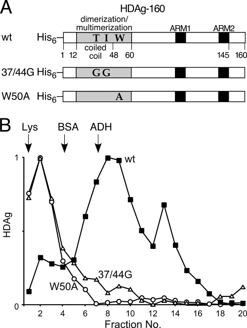
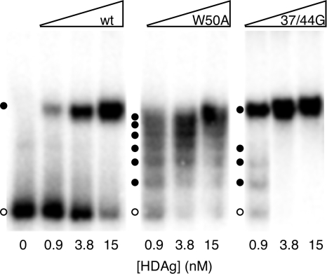
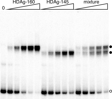
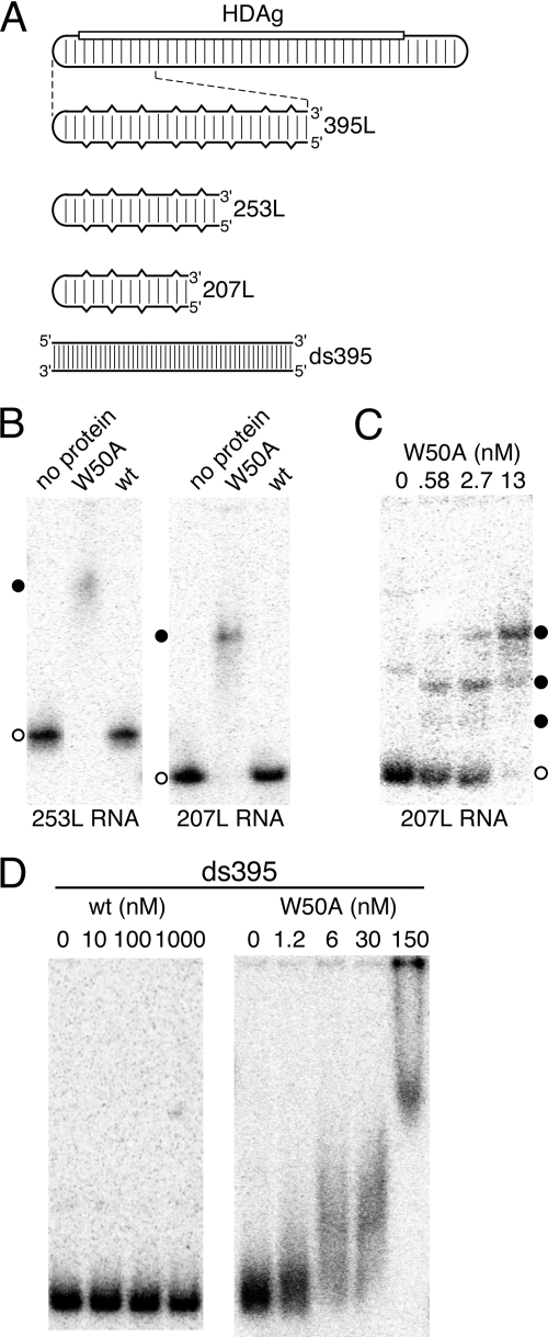
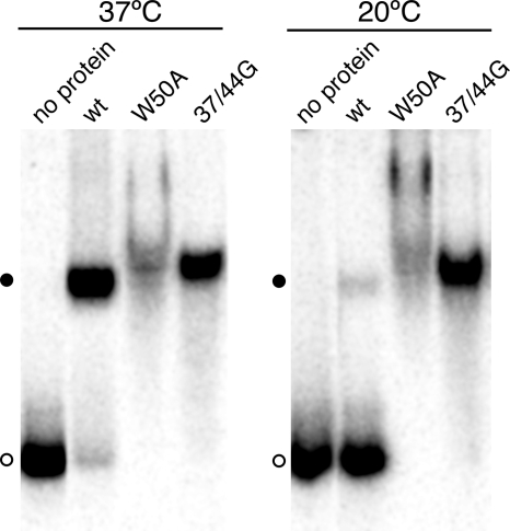
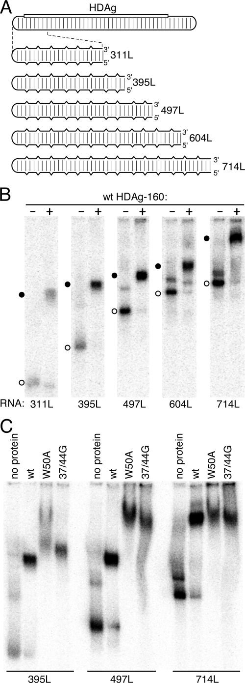
Hepatitis delta virus (HDV) RNA forms an unbranched rod structure that is associated with hepatitis delta antigen (HDAg) in cells replicating HDV. Previous in vitro binding experiments using bacterially expressed HDAg showed that the formation of a minimal ribonucleoprotein complex requires an HDV unbranched rod RNA of at least about 300 nucleotides (nt) and suggested that HDAg binds the RNA as a multimer of fixed size. The present study specifically examines the role of HDAg multimerization in the formation of the HDV ribonucleoprotein complex (RNP). Disruption of HDAg multimerization by site-directed mutagenesis was found to profoundly alter the nature of RNP formation. Mutant HDAg proteins defective for multimerization exhibited neither the 300-nt RNA size requirement for binding nor specificity for the unbranched rod structure. The results unambiguously demonstrate that HDAg binds HDV RNA as a multimer and that the HDAg multimer is formed prior to binding the RNA. RNP formation was found to be temperature dependent, which is consistent with conformational changes occurring on binding. Finally, analysis of RNPs constructed with unbranched rod RNAs successively longer than the minimum length indicated that multimeric binding is not limited to the first HDAg bound and that a minimum RNA length of between 604 and 714 nt is required for binding of a second multimer. The results confirm the previous proposal that HDAg binds as a large multimer and demonstrate that the multimer is a critical determinant of the structure of the HDV RNP.


Figures

FIG. 1.
FIG. 1.
(A) Schematic of bacterially expressed HDAg proteins. All proteins were expressed with N-terminal His6 tags. The protein multimerization region (32, 35) is shaded gray; the two arginine-rich motifs (ARM1 and ARM2) involved in RNA binding are shaded black. The amino acid identities of positions 37, 44, and 50 in the wild-type (wt) isolate used in the present study are indicated, as are the identities of these positions in the 37/44G and W50A mutants. (B) Rate zonal sucrose gradient centrifugation of bacterially expressed HDAg-160. Samples were prepared and centrifuged as described in Materials and Methods. Fraction numbers increase from the top to the bottom of the gradient. Arrows indicate the positions of peak fractions of the protein standards (Lys, lysozyme, 14.3 kDa; BSA, bovine serum albumin, 66 kDa; ADH, yeast alcohol dehydrogenase, 150 kDa).
FIG. 2.
FIG. 2.
Electrophoretic mobility shift of an HDV RNA segment with multimerization-defective HDAg mutants. 395L RNA (10 pM) was incubated with indicated concentrations of wild type (wt), W50A, or 37/44G HDAg-160 for 1 h at 37°C prior to electrophoresis in a 6% native acrylamide gel for 2 h. Open circles denote the locations of unbound RNA; solid circles indicate the locations of RNP complexes.
FIG. 3.
FIG. 3.
HDAg binds an HDV RNA segment as a preformed multimer. 395L RNA was incubated with increasing concentrations of either HDAg-160 (left lanes), HDAg-145 (middle lanes), or a mixture of equal amounts of the two proteins. The total protein concentrations were 0.5, 1.7, 6.1, 23, and 84 nM. RNP complexes formed were electrophoresed on a 6% native polyacrylamide gel for 3.5 h. The open circle denotes the location of unbound RNA; the solid circles indicate the locations of RNP complexes formed by the two proteins.
FIG. 4.
FIG. 4.
HDAg RNA binding specificity is related to protein multimerization. (A) Schematic of the 1,679-nt HDV RNA genome and RNAs analyzed for binding. The rounded rectangular lines represent HDV RNA. The location of the HDAg gene is indicated by the open rectangle. Regularly spaced vertical lines indicate 70% base-pairing between the coding and noncoding portions of the genome. In the expanded diagrams below, the partial base-pairing is indicated schematically by vertical breaks in the horizontal lines. The bottom schematic, with more closely spaced vertical lines, indicates that the RNA ds395, derived from the coding portion of the HDV RNA present in the RNA 395L, is completely double stranded. RNAs are named according to nucleotide length (8). (B to D) Native polyacrylamide gel electrophoresis after incubation of indicated 32P-labeled RNAs with the indicated concentrations of either wild-type (wt) HDAg-160 or the W50A mutant protein. Incubations were 1 h at 37°C. Protein concentrations in panel B were 30 nM. In panels B and C, open and filled circles indicate free RNA and RNPs, respectively.
FIG. 5.
FIG. 5.
Temperature dependence of binding wild-type (wt) HDAg to HDV RNA. 32P-labeled 395L RNA was incubated with 30 nM HDAg-160 (wild type, W50A, or 37/44G mutant, as indicated) for 1 h at either 20°C or 37°C, as indicated. Samples were electrophoresed for 2 h at room temperature.
FIG. 6.
FIG. 6.
HDAg binds HDV unbranched rod RNA in discrete multimeric units depending on the RNA length. (A) Schematic of the HDV RNA genome and the series of RNAs analyzed for binding, labeled as in Fig. 4. (B and C) Native polyacrylamide gel electrophoresis after incubation of the indicated 32P-labeled RNAs (20 pM) with either no protein or 30 nM HDAg-160 (wild type, W50A mutant, or 37/44G mutant, as indicated). Circles indicate unbound RNA (open) or RNP complexes (filled). In panel B, the time of electrophoresis was 3 h. In panel C, the electrophoresis times were varied: 395L, 2.65 h; 497L, 3.1 h; and 714L, 4 h.

Similar articles

Cited by

References

    1. Antson, A. A., E. J. Dodson, G. Dodson, R. B. Greaves, X. Chen, and P. Gollnick. 1999. Structure of the trp RNA-binding attenuation protein, TRAP, bound to RNA. Nature 401:235-242. - PubMed
    1. Baumann, C., J. Otridge, and P. Gollnick. 1996. Kinetic and thermodynamic analysis of the interaction between TRAP (trp RNA-binding attenuation protein) of Bacillus subtilis and trp leader RNA. J. Biol. Chem. 271:12269-12274. - PubMed
    1. Bichko, V. V., S. M. Lemon, J. G. Wang, S. Hwang, M. M. Lai, and J. M. Taylor. 1996. Epitopes exposed on hepatitis delta virus ribonucleoproteins. J. Virol. 70:5807-5811. - PMC - PubMed
    1. Casey, J. L., and J. L. Gerin. 1998. Genotype-specific complementation of hepatitis delta virus RNA replication by hepatitis delta antigen. J. Virol. 72:2806-2814. - PMC - PubMed
    1. Chang, J., X. Nie, H. E. Chang, Z. Han, and J. Taylor. 2008. Transcription of hepatitis delta virus RNA by RNA polymerase II. J. Virol. 82:1118-1127. - PMC - PubMed

Publication types

MeSH terms