. 2010 Feb;84(3):1574-84.
doi: 10.1128/JVI.01326-09. Epub 2009 Nov 18.

Porcine reproductive and respiratory syndrome virus nonstructural protein 1beta modulates host innate immune response by antagonizing IRF3 activation

Affiliations

Porcine reproductive and respiratory syndrome virus nonstructural protein 1beta modulates host innate immune response by antagonizing IRF3 activation

Lalit K Beura et al. J Virol. 2010 Feb.

Abstract

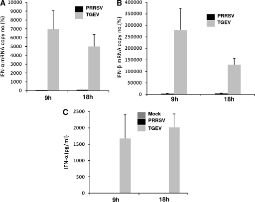
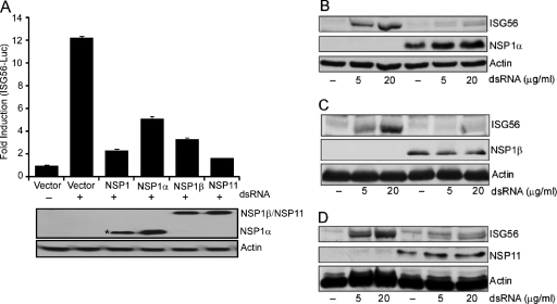
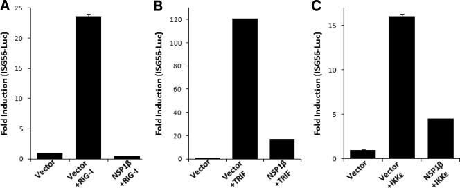
Porcine reproductive and respiratory syndrome virus (PRRSV) infection of swine leads to a serious disease characterized by a delayed and defective adaptive immune response. It is hypothesized that a suboptimal innate immune response is responsible for the disease pathogenesis. In the study presented here we tested this hypothesis and identified several nonstructural proteins (NSPs) with innate immune evasion properties encoded by the PRRS viral genome. Four of the total ten PRRSV NSPs tested were found to have strong to moderate inhibitory effects on beta interferon (IFN-beta) promoter activation. The strongest inhibitory effect was exhibited by NSP1 followed by, NSP2, NSP11, and NSP4. We focused on NSP1alpha and NSP1beta (self-cleavage products of NSP1 during virus infection) and NSP11, three NSPs with strong inhibitory activity. All of three proteins, when expressed stably in cell lines, strongly inhibited double-stranded RNA (dsRNA) signaling pathways. NSP1beta was found to inhibit both IFN regulatory factor 3 (IRF3)- and NF-kappaB-dependent gene induction by dsRNA and Sendai virus. Mechanistically, the dsRNA-induced phosphorylation and nuclear translocation of IRF3 were strongly inhibited by NSP1beta. Moreover, when tested in a porcine myelomonocytic cell line, NSP1beta inhibited Sendai virus-mediated activation of porcine IFN-beta promoter activity. We propose that this NSP1beta-mediated subversion of the host innate immune response plays an important role in PRRSV pathogenesis.


Figures

FIG. 1.
FIG. 1.
Inhibition of type I IFN production after PRRSV infection. Porcine monocyte-derived macrophages were mock infected or infected with PRRSV (FL-12) or TGEV at a multiplicity of infection of 1. Supernatants and cells were collected after 9 and 18 h p.i., respectively. Total RNA isolated from cells was reverse transcribed, and real-time PCR was performed for the detection of porcine IFN-α (A) and IFN-β (B) mRNA. The mRNA copy numbers were calculated after normalization with the β-actin copy number and are expressed as percentages relative to the mock control. Bars show the average of mRNA copy numbers ± the standard error of the mean (SEM) from three independent experiments using cells isolated from three different pigs. (C) Supernatants from the same experiments were used to detect secreted IFN-α by ELISA. Quantified recombinant porcine IFN-α was used as a standard, and IFN-α levels were calculated based upon a standard curve.
FIG. 2.
FIG. 2.
Specific PRRSV proteins are involved in downregulation of IFN-β promoter. (A) HeLa cells were cotransfected with pIFN-β-CAT plasmid (1 μg), IRF3 expression plasmid (0.5 μg), and the viral nonstructural protein expression plasmids (1 μg) or bovine herpesvirus type 1 protein bICP0. Empty vector was added to the transfection mixture to keep the total DNA amount constant. Cell extracts collected at 40 h posttransfection were analyzed for CAT activity as described in Materials and Methods. The value of the CAT activity in the presence of empty vector was set at 100%, and those of the viral proteins were normalized accordingly. (B) Increasing amounts of indicated NSP coding plasmids were transfected, and CAT activities were measured as described earlier. The bars represent the means ± the SEM from three independent experiments. The bottom panels show increasing levels of individual NSP expression upon transfection (actin was used as a loading control).
FIG. 3.
FIG. 3.
Inhibition of IRF3-mediated gene induction by PRRSV proteins. (A) HEK-293TLR3 cells were cotransfected with the indicated viral NSP expression plasmids (1 μg) or empty vector, ISG56-luciferase plasmid (0.4 μg), and pRLTK (0.01 μg). At 40 h posttransfection the cells were treated with 10 μg of dsRNA/ml or PBS for 6 h and assayed for luciferase activity. The bars represent the average fold inductions ± the SEM for luciferase activities compared to PBS-treated vector control. The panel below the bar graph shows the expression of the respective NSPs. The asterisk (*) at NSP1 lane indicates the detection of proteolytically processed NSP1 to NSP1α, which carries the N-terminal FLAG tag. (B to D) HEK293-TLR3 cells stably expressing NSP1α (B), NSP1β (C), and NSP11 (D) (lanes 4 to 6) were stimulated with the indicated amount of dsRNA for 6 h, and the ISG56 level was measured by immunoblotting and compared to empty vector-transfected, puromycin-resistant control cells (lanes 1 to 3). Expressions of NSPs were detected by probing with anti-FLAG/HA antibody.
FIG. 4.
FIG. 4.
Inhibition of NF-κB-mediated gene induction by PRRSV proteins. (A) HEK293-TLR3 cells were cotransfected with the indicated viral NSP expression plasmids or empty vector (1 μg), 5X NF-κB-luciferase plasmid (0.5 μg), and pRLTK (0.025 μg). At 40 h posttransfection the cells were treated with dsRNA for 6 h and assayed for luciferase activity. The fold induction of the average luciferase activity compared to the untreated vector control from a representative experiment is shown. The immunoblot panel indicates the expression of individual NSPs in the luciferase assay. (B) IL-8 mRNA levels were measured by real-time PCR in HEK293-TLR3 cells stably expressing NSP1β or vector control cells with or without dsRNA treatment. The mRNA copy numbers were calculated after normalizing them with the RPL32 (internal control) copy number and are expressed as percentages relative to the uninduced vector control.
FIG. 5.
FIG. 5.
NSP1β inhibits IRF3 signaling by acting downstream of IRF3 kinases. HEK293-TLR3 cells were cotransfected with ISG56-luciferase plasmid, NSP1β expression plasmids, or empty vector. IRF3 signaling pathway was induced by including in the transfection constitutively active RIG-I (A), TRIF (B), and IKKɛ (C). Cells were lysed 40 h posttransfection and assayed for luciferase activity. Bars indicate the mean fold inductions ± the SEM of luciferase activity compared to the vector control from three independent experiments.
FIG. 6.
FIG. 6.
NSP1β inhibits dsRNA-induced IRF3 phosphorylation and nuclear translocation. (A) HEK293-TLR3 cells stably expressing NSP1β- or vector-transfected cells (vector) were either mock treated or treated with dsRNA for 2 h. Phosphorylated IRF3 (pIRF3) and total IRF3 levels were measured by immunoblotting. (B) Nuclear fractions (N.P) from HEK293-TLR3 cells stably expressing NSP1β either mock treated or treated with dsRNA were blotted for IRF3. DRBP76 and tubulin are markers for nuclear and cytoplasmic fractions, respectively. WCE, whole-cell extract. (C) IRF3 subcellular localization was determined by confocal microscopy in HEK-293TLR3 cells stably transfected with empty vector (vector) or NSP1β after dsRNA stimulation. For this purpose, cells were either treated or left untreated with 100 μg of dsRNA/ml for 2 h, followed by immunofluorescence for endogenous IRF3 (green) and NSP1β (red) with rabbit anti-IRF3 and mouse anti-HA antibody, as indicated at the top of the panel. Position of nucleus is indicated by DAPI (blue) staining in the merge image.
FIG. 7.
FIG. 7.
NSP1 and NSP1β inhibit SeV-induced porcine IFN-β promoter activation and human ISG56 induction. (A) HEK293-TLR3 cells stably transfected with empty vector (vector) or NSP1β were either mock infected or infected with SeV (40 HA units/ml) for 8 h. Endogenous ISG56 levels were detected by immunoblotting. NSP1β protein was detected by anti-HA antibody. (B) Porcine monocytic cells (3D4/31 cell line) were cotransfected with increasing concentrations of NSP1/NSP1β expression plasmids or empty vector (vector), along with porcine IFN-β-luciferase plasmid (0.4 μg) and pRLTK (0.01 μg). At 40 h after the transfection cells were either mock infected or infected with 80 HA units of SeV/ml for 8 h and assayed for luciferase activity. Bars indicate the average fold induction of luciferase activities ± the SEM compared to uninfected vector control cells. The bottom panel shows immunoblots for the level of each NSP expression in respective transfection. The asterisk indicates detection of N-terminally FLAG-tagged NSP1α, the cleavage product of NSP1.

References

    1. Albina, E., C. Carrat, and B. Charley. 1998. Interferon-alpha response to swine arterivirus (PoAV), the porcine reproductive and respiratory syndrome virus. J. Interferon Cytokine Res. 18:485-490. - PubMed
    1. Allende, R., W. W. Laegreid, G. F. Kutish, J. A. Galeota, R. W. Wills, and F. A. Osorio. 2000. Porcine reproductive and respiratory syndrome virus: description of persistence in individual pigs upon experimental infection. J. Virol. 74:10834-10837. - PMC - PubMed
    1. Ansari, I. H., B. Kwon, F. A. Osorio, and A. K. Pattnaik. 2006. Influence of N-linked glycosylation of porcine reproductive and respiratory syndrome virus GP5 on virus infectivity, antigenicity, and ability to induce neutralizing antibodies. J. Virol. 80:3994-4004. - PMC - PubMed
    1. Bauhofer, O., A. Summerfield, Y. Sakoda, J. D. Tratschin, M. A. Hofmann, and N. Ruggli. 2007. Classical swine fever virus Npro interacts with interferon regulatory factor 3 and induces its proteasomal degradation. J. Virol. 81:3087-3096. - PMC - PubMed
    1. Bautista, E. M., K. S. Faaberg, D. Mickelson, and E. D. McGruder. 2002. Functional properties of the predicted helicase of porcine reproductive and respiratory syndrome virus. Virology 298:258-270. - PMC - PubMed

Publication types

MeSH terms

LinkOut - more resources