. 2010 Jan 22;285(4):2415-27.
doi: 10.1074/jbc.M109.071324. Epub 2009 Nov 18.

Structure of a eukaryotic nonribosomal peptide synthetase adenylation domain that activates a large hydroxamate amino acid in siderophore biosynthesis

Affiliations

Structure of a eukaryotic nonribosomal peptide synthetase adenylation domain that activates a large hydroxamate amino acid in siderophore biosynthesis

T Verne Lee et al. J Biol Chem. .

Abstract

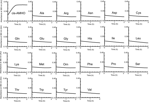
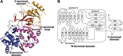
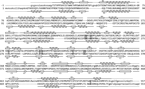
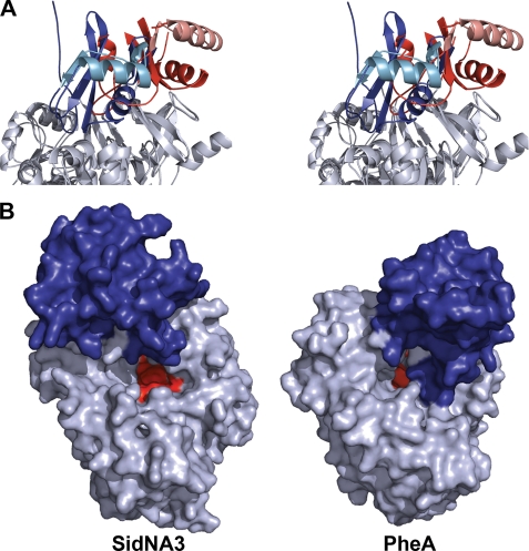
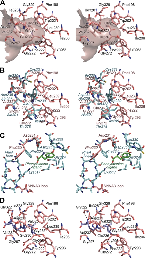
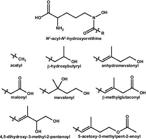
Nonribosomal peptide synthetases (NRPSs) are large, multidomain proteins that are involved in the biosynthesis of an array of secondary metabolites. We report the structure of the third adenylation domain from the siderophore-synthesizing NRPS, SidN, from the endophytic fungus Neotyphodium lolii. This is the first structure of a eukaryotic NRPS domain, and it reveals a large binding pocket required to accommodate the unusual amino acid substrate, N(delta)-cis-anhydromevalonyl-N(delta)-hydroxy-L-ornithine (cis-AMHO). The specific activation of cis-AMHO was confirmed biochemically, and an AMHO moiety was unambiguously identified as a component of the fungal siderophore using mass spectroscopy. The protein structure shows that the substrate binding pocket is defined by 17 amino acid residues, in contrast to both prokaryotic adenylation domains and to previous predictions based on modeling. Existing substrate prediction methods for NRPS adenylation domains fail for domains from eukaryotes due to the divergence of their signature sequences from those of prokaryotes. Thus, this new structure will provide a basis for improving prediction methods for eukaryotic NRPS enzymes that play important and diverse roles in the biology of fungi.


Figures

FIGURE 1.
FIGURE 1.
SidNA3 activity assays. Indicative plots of the accumulation of PPi, as monitored by absorbance at 360 nm, for SidNA3 activity using the 20 proteogenic amino acids, cis-AMHO, and ornithine (Orn) as substrates. The assay was conducted three times using different concentrations of enzyme and substrate. In each case, cis-AMHO was the only amino acid that was adenylated. Subsequently, duplicate assays were conducted to calculate observed kcat and Km values (see text for details). AU, absorbance units.
FIGURE 2.
FIGURE 2.
Structure and topology of SidNA3. A, ribbon diagram of the SidNA3 structure. The N-terminal domain is made up of a distorted six-stranded antiparallel β-barrel (red) together with an αβαβα structure formed by two predominantly parallel β-sheets flanked by α-helices (blue and purple). The C-terminal domain (orange) is made up of a three-stranded antiparallel β-sheet surrounded by three α-helices. A long loop in the C-terminal domain, located between the final β-strand and the final α-helix, loops back toward the N-terminal domain and is marked. B, topology diagram of SidNA3. Diagram is drawn in the same style as that of the PheA topology diagram in Conti et al. (9) and using the same numbering scheme for the β-strands. The circles represent α-helices and the arrows β-strands. The locations of the chain breaks (because of poor electron density) in the SidNA3 model are marked.
FIGURE 3.
FIGURE 3.
Structure-based sequence alignment. Structure-based sequence alignment of the protein sequences SidNA3 (top) and PheA (bottom) (9). The positions of the β-strands and α-helices for both structures are shown. The numbering scheme for the β-strands is that used in Conti et al. (9). The residues not present in the models are indicated by lowercase letters, and the first five N-terminal residues of SidNA3, which are cloning artifacts, are shown in gray lettering and given negative numbers. The residues involved in binding the amino acid substrate are shaded (dark gray for the standard PheA residues and light gray for the extra SidNA3 residues).
FIGURE 4.
FIGURE 4.
Orientation of the C-terminal domain for SidNA3. A, stereo ribbon diagram showing the orientation of the C-terminal domain in SidNA3 compared with the adenylation conformation of PheA (9). The C-terminal domain of SidNA3 is red and that of PheA is blue. The N-terminal domains have been aligned and are shown in gray. The second α-helix and subsequent β-strand of the C-terminal domains are colored in lighter shades to aid visualization of the rotation between the conformations. B, molecular surface diagram showing the accessibility of the active sites in the open conformation of SidNA3 compared with the adenylation conformation of PheA (9). The amino acid binding pocket is shown in red, and the C-terminal domain is colored blue.
FIGURE 5.
FIGURE 5.
Amino acid binding pocket of SidNA3. A, stereo “Connolly” surface (48) diagram of the amino acid binding pocket of SidNA3 and the residues lining it. The entrance to the pocket is to the left of the diagram. B, stereo diagram comparing the residues lining the amino acid binding pockets of SidNA3 and PheA (9). The SidNA3 residues are pink, and the PheA residues are blue. The residues of PheA are labeled in blue italics. C, stereo diagram comparing residues involved in binding the main chain atoms of the amino acid ligand in SidNA3 and PheA (9). The SidNA3 residues are pink; the PheA residues are blue, and the phenylalanine ligand of PheA is green. The C-terminal domain loops of both molecules are also shown. The only binding pocket-lining residues shown are those of PheA. The hydrogen bonds between PheA and the phenylalanine ligand are shown. D, stereo diagram of cis-AMHO docked into the binding pocket of SidNA3. The SidNA3 residues lining the binding pocket are pink. The top-ranked solution for the docking of the cis-AMHO ligand is shown in dark blue. The hydrogen bonds between SidNA3 and the docked ligand are shown (a hydrogen bond between the α-amino group of the ligand and Asp-231 is hidden behind the ligand).
FIGURE 6.
FIGURE 6.
Nδ-Acyl-Nδ-hydroxy-l-ornithine amino acids found in fungal hydroxamate siderophores. The various R groups are shown below the parent at the top of the figure.

References

    1. Finking R., Marahiel M. A. (2004) Annu. Rev. Microbiol. 58, 453–488 - PubMed
    1. Fleetwood D. J., Scott B., Lane G. A., Tanaka A., Johnson R. D. (2007) Appl. Environ. Microbiol. 73, 2571–2579 - PMC - PubMed
    1. Sieber S. A., Marahiel M. A. (2005) Chem. Rev. 105, 715–738 - PubMed
    1. Murzin A. G., Brenner S. E., Hubbard T., Chothia C. (1995) J. Mol. Biol. 247, 536–540 - PubMed
    1. Finn R. D., Tate J., Mistry J., Coggill P. C., Sammut S. J., Hotz H. R., Ceric G., Forslund K., Eddy S. R., Sonnhammer E. L., Bateman A. (2008) Nucleic Acids Res. 36, D281–D288 - PMC - PubMed

Publication types

Associated data

LinkOut - more resources