. 2010 Jan 15;285(3):1773-80.
doi: 10.1074/jbc.M109.067470. Epub 2009 Nov 18.

Crystal structures of Trypanosoma brucei sterol 14alpha-demethylase and implications for selective treatment of human infections

Affiliations

Crystal structures of Trypanosoma brucei sterol 14alpha-demethylase and implications for selective treatment of human infections

Galina I Lepesheva et al. J Biol Chem. .

Abstract

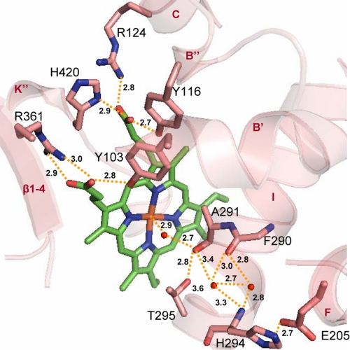
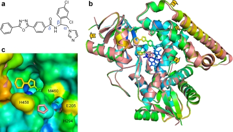
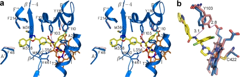
Sterol 14alpha-demethylase (14DM, the CYP51 family of cytochrome P450) is an essential enzyme in sterol biosynthesis in eukaryotes. It serves as a major drug target for fungal diseases and can potentially become a target for treatment of human infections with protozoa. Here we present 1.9 A resolution crystal structures of 14DM from the protozoan pathogen Trypanosoma brucei, ligand-free and complexed with a strong chemically selected inhibitor N-1-(2,4-dichlorophenyl)-2-(1H-imidazol-1-yl)ethyl)-4-(5-phenyl-1,3,4-oxadi-azol-2-yl)benzamide that we previously found to produce potent antiparasitic effects in Trypanosomatidae. This is the first structure of a eukaryotic microsomal 14DM that acts on sterol biosynthesis, and it differs profoundly from that of the water-soluble CYP51 family member from Mycobacterium tuberculosis, both in organization of the active site cavity and in the substrate access channel location. Inhibitor binding does not cause large scale conformational rearrangements, yet induces unanticipated local alterations in the active site, including formation of a hydrogen bond network that connects, via the inhibitor amide group fragment, two remote functionally essential protein segments and alters the heme environment. The inhibitor binding mode provides a possible explanation for both its functionally irreversible effect on the enzyme activity and its selectivity toward the 14DM from human pathogens versus the human 14DM ortholog. The structures shed new light on 14DM functional conservation and open an excellent opportunity for directed design of novel antiparasitic drugs.


Figures

FIGURE 1.
FIGURE 1.
Ligand-free Tbb14DM (salmon). a, superposition with MtCYP51 (1e9x) (green), ribbon diagram. The heme, Tyr-116 (Tbb), and Phe-89 (Mt) are shown in stick representation. The most divergent active site-defining regions are indicated with black arrows, and distances between the equivalent Cα atoms are provided in parentheses. The locations of the putative substrate entrance in Tbb and Mt structures are shown with red and green arrows, respectively. The structure of ligand-free MtCYP51 (1h5z) is identical to 1e9x except for its larger opening to the upper surface due to missing density for helix C. Left inset, I-helix in Tbb and Mt CYP51, distal view. Right inset, BC loop, upper view. b, surface representations of Tbb and Mt CYP51 from the upper and distal sides; the corresponding heme orientation is shown on the right. Inside the proteins, the heme is presented as spheres. In Tbb14DM, the channel extends about 20 Å from the distal surface to the iron, forming an angle of about 50° to the heme plane. In MTCYP51, the funnel-like opening runs from above perpendicular to the heme plane.
FIGURE 2.
FIGURE 2.
Heme environment in ligand-free Tbb14DM. Heme and surrounding residues are shown in stick representation, H-bonds are indicated with yellow dashes, and the distances (Å) are marked. Upper region, residues interacting with the heme propionates. The A-ring forms H-bonds with Tyr-103 (helix B′) and Arg-361 (β1–4), and the D-ring is supported by Tyr-116 (helix B″), Arg-124 (helix C), and His-420 (helix K″). Lower region, potential I-helix-based proton delivery route. The water molecules included into the H-bond network are presented as small red spheres. The I-helix groove is formed by protrusion of the Ala-291 carbonyl toward the heme and probably modulated by flexible Gly-292. As a result, two helical H-bonds (Phe-290–His-294 and Ala-291–Thr-295) are missing, the peptide carbonyl of Phe-290 being connected through the waters. Ala-291, Gly-292, His-294, and Thr-295 (“the conserved threonine” in the majority of CYP families) are part of the CYP51 I-helix signature (–aGqHTS–). His-294 is the residue specific for the 14DM; the other P450s usually have Asp/Glu in this position. Glu-205 (helix F), which forms a salt bridge with His-294, is also conserved in all 14DM.
FIGURE 3.
FIGURE 3.
VNI-bound Tbb14DM. a, the chemical structure of VNI. b, superposition of the ligand-free (salmon) and VNI-bound (colored by B-factor) Tbb14DM structures, distal view. The B-factor ranges from blue minimum to red maximum within 17–68 Å2. The heme, VNI, and three residues surrounding the entrance (Ile-45, Pro-210, and His-458) are presented as stick models. Four structural segments where the backbone in all four molecules moves >0.5 Å away from the catalytic core relative to the ligand-free structure are marked with arrows showing the directions of coordinate shifts. 1, N terminus (residues 32–65); 2, BC segment (95–114); 3, FG segment (185–232); and 4, middle part of helix I (288–292). Superimposition of four ligand-free and four VNI-bound molecules can be seen in supplemental Fig. S5. c, enlarged view of the access channel in surface representation; orientation is the same as in b. Some of the heme atoms are seen as blue spheres. The salmon ribbon of the ligand-free structure is partially visible where the shift in the backbone is larger than 1 Å (helices A, F″, and I). The second channel is bordered by helices I (His-294), F (Glu-205), and β4 hairpin (Met-460).
FIGURE 4.
FIGURE 4.
Active site of Tbb14DM with bound VNI. a, secondary structure and stick representations of the 15 residues (blue) within 4 Å from VNI (yellow) shown in stereo view. Location of the corresponding side chains in the ligand-free structure, if different, can be seen as pink lines. The hydrogen bonds around the VNI amide group fragment are indicated as purple dashes. b, altered location of Tyr-103 in ligand-free (salmon) and VNI-bound (blue) Tbb14DM.
FIGURE 5.
FIGURE 5.
14DM in T. brucei. a and b, immunodetection of 14DM in Tbb procyclic (PF) and bloodstream forms (BF). a, whole cell extracts of procyclic and mice-isolated bloodstream form trypanosomes were probed by Western blotting with mouse anti-Tbb14DM antiserum. Recombinant (r) Tbb14DM served as positive control. b, mice-isolated bloodstream form cell extracts (10 mg) were immunoprecipitated with rat anti-Tbb14DM antiserum. The immunoprecipitated material (IP) was analyzed by Western blotting after non-reducing electrophoresis with mouse polyclonal anti-Tbb14DM antiserum. Controls were performed in the absence of either cell extracts or rat antiserum. Tbb14DM served as positive control. c, suppression of Tbb infection in mice by oral administration of VNI (20 mg/kg) or clotrimazole (CLT, 50 and 100 mg/kg). Clotrimazole was selected among several commercially available antifungal azoles by testing their inhibitory potency in reconstituted Tbb14DM reaction in vitro due to the limited amount of VNI. The differences observed between the mean of treated and mock-treated mice are statistically significant as assessed by Student's t test (p < 0.01). Error bars indicate S.E.

Similar articles

Cited by

References

    1. Barrett M. P., Burchmore R. J., Stich A., Lazzari J. O., Frasch A. C., Cazzulo J. J., Krishna S. (2003) Lancet 362, 1469–1480 - PubMed
    1. Croft S. L., Barrett M. P., Urbina J. A. (2005) Trends Parasitol. 21, 508–512 - PubMed
    1. Myler P. J. (2008) Adv. Exp. Med. Biol. 625, 133–140 - PMC - PubMed
    1. Soeiro M. N., de Castro S. L. (2009) Expert. Opin. Ther. Targets 13, 105–121 - PubMed
    1. Lüscher A., de Koning H. P., Mäser P. (2007) Curr. Pharm. Des. 13, 555–567 - PubMed

Publication types

MeSH terms