. 2010 Jan 15;285(3):1671-80.
doi: 10.1074/jbc.M109.061630. Epub 2009 Nov 18.

A novel epimerase that converts GlcNAc-P-P-undecaprenol to GalNAc-P-P-undecaprenol in Escherichia coli O157

Affiliations

A novel epimerase that converts GlcNAc-P-P-undecaprenol to GalNAc-P-P-undecaprenol in Escherichia coli O157

Jeffrey S Rush et al. J Biol Chem. .

Abstract

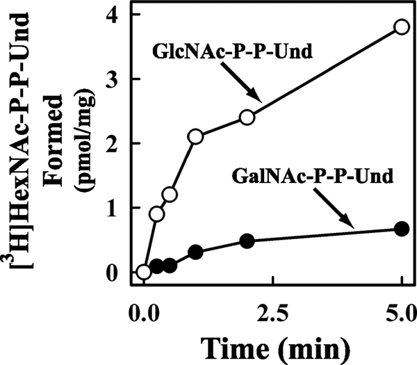
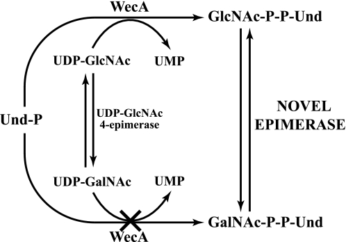
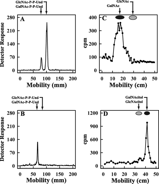
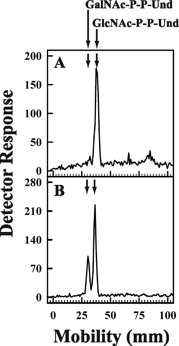
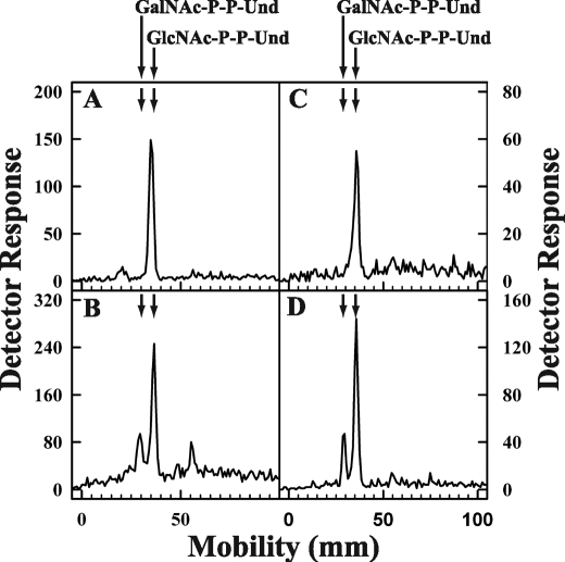
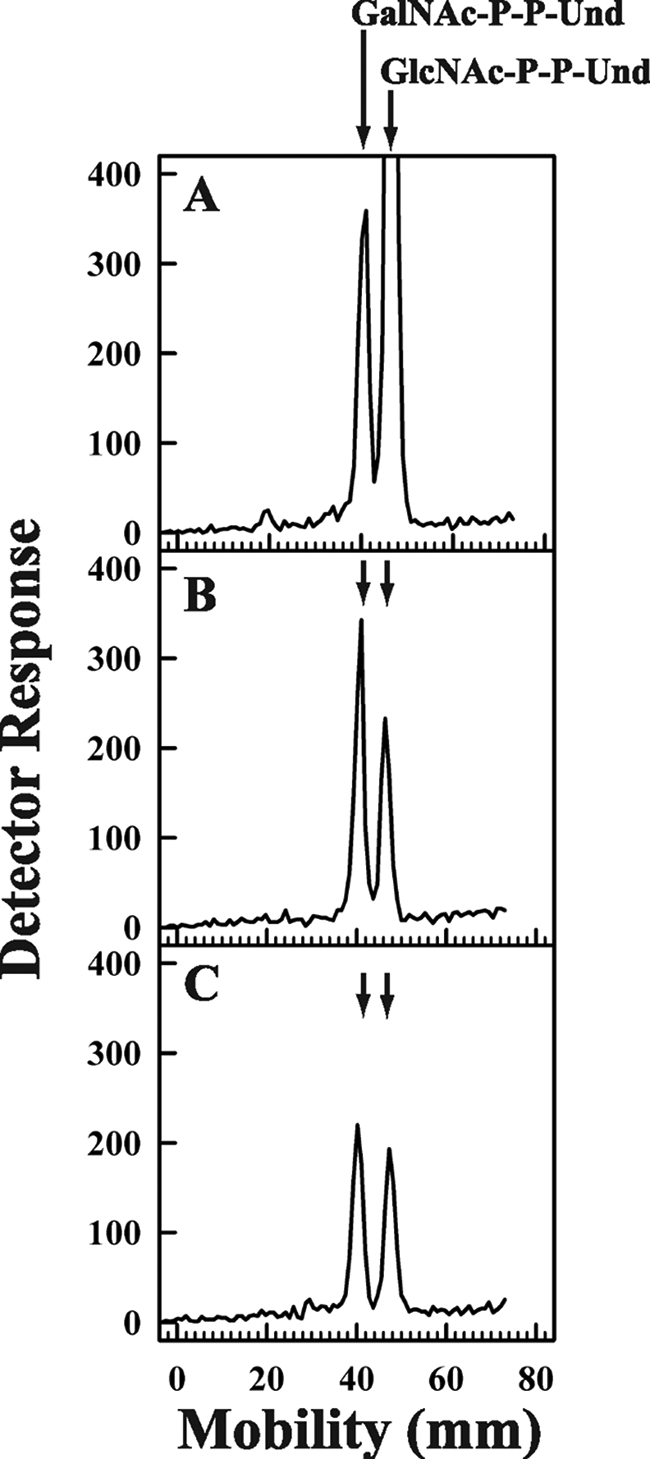
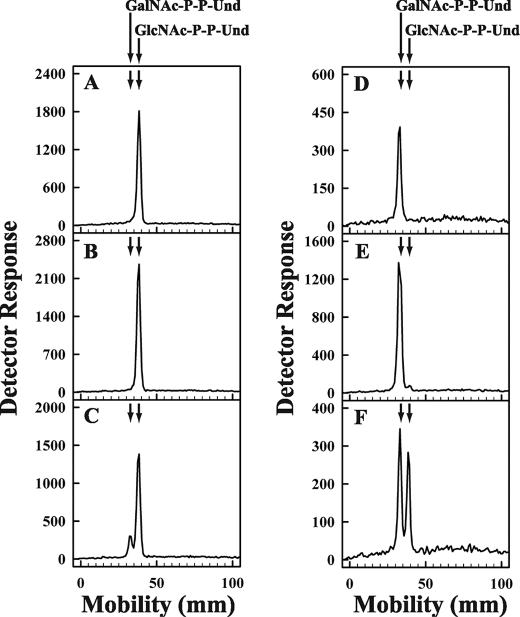
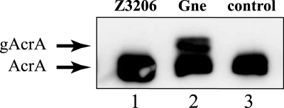
Escherichia coli strain O157 produces an O-antigen with the repeating tetrasaccharide unit alpha-D-PerNAc-alpha-l-Fuc-beta-D-Glc-alpha-D-GalNAc, preassembled on undecaprenyl pyrophosphate (Und-P-P). These studies were conducted to determine whether the biosynthesis of the lipid-linked repeating tetrasaccharide was initiated by the formation of GalNAc-P-P-Und by WecA. When membrane fractions from E. coli strains K12, O157, and PR4019, a WecA-overexpressing strain, were incubated with UDP-[3H]GalNAc, neither the enzymatic synthesis of [3H]GlcNAc-P-P-Und nor [3H]GalNAc-P-P-Und was detected. However, when membrane fractions from strain O157 were incubated with UDP-[3H]GlcNAc, two enzymatically labeled products were observed with the chemical and chromatographic properties of [3H]GlcNAc-P-P-Und and [3H]GalNAc-P-P-Und, suggesting that strain O157 contained an epimerase capable of interconverting GlcNAc-P-P-Und and GalNAc-P-P-Und. The presence of a novel epimerase was demonstrated by showing that exogenous [3H]GlcNAc-P-P-Und was converted to [3H]GalNAc-P-P-Und when incubated with membranes from strain O157. When strain O157 was metabolically labeled with [3H]GlcNAc, both [3H]GlcNAc-P-P-Und and [3H]GalNAc-P-P-Und were detected. Transformation of E. coli strain 21546 with the Z3206 gene enabled these cells to synthesize GalNAc-P-P-Und in vivo and in vitro. The reversibility of the epimerase reaction was demonstrated by showing that [3H]GlcNAc-P-P-Und was reformed when membranes from strain O157 were incubated with exogenous [3H]GalNAc-P-P-Und. The inability of Z3206 to complement the loss of the gne gene in the expression of the Campylobacter jejuni N-glycosylation system in E. coli indicated that it does not function as a UDP-GlcNAc/UDP-GalNAc epimerase. Based on these results, GalNAc-P-P-Und is synthesized reversibly by a novel GlcNAc-P-P-Und epimerase after the formation of GlcNAc-P-P-Und by WecA in E. coli O157.


Figures

FIGURE 1.
FIGURE 1.
Time course of [3H]GlcNAc/GalNAc-P-P-Und synthesis by membrane fractions from E. coli O157. The membrane fraction from E. coli strain O157 was incubated with UDP-[3H]GlcNAc for the indicated times at 37 °C. The [3H]lipid products were extracted, and the incorporation of [3H]GlcNAc into [3H]GlcNAc-P-P-Und (○) and [3H]GalNAc-P-P-Und (●) was assayed as described under “Experimental Procedures.”
FIGURE 2.
FIGURE 2.
Proposed biosynthetic pathway for the formation of GalNAc-P-P-Und from GlcNAc-P-P-Und.
FIGURE 3.
FIGURE 3.
Purification and Characterization of [3H]GalNAc-P-P-Und synthesized by membrane fractions from E. coli strain O157. Membrane fractions from E. coli O157 were incubated with UDP-[3H]GlcNAc, and the [3H]GalNAc lipids were purified as described under “Experimental Procedures.” Panel A, preparative thin layer chromatogram of [3H]HexNAc lipids on borate-impregnated silica gel G (Quantum 1) after purification on DEAE-cellulose is shown. Panel B, thin layer chromatography of purified [3H]GalNAc-P-P-Und on borate-impregnated silica gel G (Baker, Si250) after recovery from the preparative plate in panel A is shown. Panel C, descending paper chromatogram (borate-impregnated Whatman No. 1 paper) of the 3H amino sugar recovered after mild acid hydrolysis of [3H]GalNAc-P-P-Und purified in panel B is shown. Panel D, descending paper chromatogram (Whatman No. 3MM) of the [3H]HexNAc-alditol produced by reduction of the [3H] amino sugar from panel C with NaBH4.
FIGURE 4.
FIGURE 4.
Metabolic labeling of E. coli 21546 cells and E. coli 21546 cells after transformation with pMLBAD:Z3206. E. coli 21546 (panel A) and E. coli 21546:pMLBAD/Z3206 (panel B) were labeled metabolically with [3H]GlcNAc for 5 min at 37 °C. [3H]GlcNAc/GalNAc-P-P-Und were extracted, freed of water soluble contaminants, and separated by thin layer chromatography on borate-impregnated silica gel plates (Baker Si250) as described under “Experimental Procedures.” Radioactive lipids were detected using a Bioscan chromatoscanner. The chromatographic positions of GalNAc-P-P-Und and GlcNAc-P-P-Und are indicated by arrows.
FIGURE 5.
FIGURE 5.
Thin layer chromatography of [3H]GlcNAc/GalNAc-P-P-Und formed by incubation of membrane fractions from E. coli strains with UDP-[3H]GlcNAc. Membrane fractions from E. coli strains K12 (panel A), O157 (panel B), 21546 (panel C), and 21546:pMLBAD/Z3206 (panel D) were incubated with UDP-[3H]GlcNAc for 10 min at 37 °C, and the [3H]lipid products were extracted, freed of water-soluble contaminants by partitioning, and separated by thin layer chromatography on borate-impregnated silica gel plates (Baker Si250) as described under “Experimental Procedures.” The chromatographic positions of GalNAc-P-P-Und and GlcNAc-P-P-Und are indicated by arrows.
FIGURE 6.
FIGURE 6.
Discharge of GlcNAc-P by incubation with UMP. Membrane fractions from E. coli 21546:Z3206 were preincubated with UDP-[3H]GlcNAc to enzymatically label GlcNAc-P-P-Und for 10 min (panel A) at 37 °C followed by a second incubation period with 1 mm UMP included for either 1 min (panel B) or 2 min (panel C). After the indicated incubation periods [3H]GlcNAc/GalNAc-P-P-Und were extracted and resolved by thin layer chromatography on borate-impregnated silica gel plates (Baker Si250) as described under “Experimental Procedures.” The chromatographic positions of GalNAc-P-P-Und and GlcNAc-P-P-Und are indicated by arrows.
FIGURE 7.
FIGURE 7.
Conversion of exogenous [3H]GlcNAc-P-P-Und and [3H]GalNAc-P-P-Und to the pertinent [3H]HexNAc-P-P-Und product catalyzed by membranes from strain 21546 expressing Z3206. Membrane fractions from E. coli strain 21546 (panels B and E) and 21546:pMLBAD/Z3206 (panels C and F) were incubated with purified [3H]GlcNAc-P-P-Und (panels A, B, and C) or [3H]GalNAc-P-P-Und (panels D, E, and F) (dispersed ultrasonically in 1% Triton X-100) for 1 min at 37 °C. [3H]GlcNAc/GalNAc-P-P-Und were extracted, resolved by thin layer chromatography on borate-impregnated silica gel plates (Baker Si250), and detected with a Bioscan AR2000 radiochromatoscanner as described under “Experimental Procedures.”
FIGURE 8.
FIGURE 8.
Z3206 does not complement glycosylation of AcrA in a Gne-dependent glycosylation system. Periplasmic extracts prepared from E. coli DH5α cells carrying the AcrA expression plasmid and the pgl operon Δgne complemented with pMLBAD:Z3206 (lane 1), pMLBAD:gne (lane 2), or the vector control pMLBAD (lane 3) were separated by 10% SDS-PAGE and transferred to nitrocellulose membranes. AcrA and its glycosylated forms were detected with anti AcrA antisera. The position of bands corresponding to unglycosylated (AcrA) and glycosylated AcrA (gAcrA) is indicated.

References

    1. Raetz C. R., Whitfield C. (2002) Annu. Rev. Biochem. 71, 635–700 - PMC - PubMed
    1. Whitfield C. (2006) Annu. Rev. Biochem. 75, 39–68 - PubMed
    1. Samuel G., Reeves P. R. (2003) Carbohydr. Res. 338, 2503–2519 - PubMed
    1. Reeves P. R., Hobbs M., Valvano M. A., Skurnik M., Whitfield C., Coplin D., Kido N., Klena J., Maskell D., Raetz C. R., Rick P. D. (1996) Trends Microbiol. 4, 495–503 - PubMed
    1. Law D. (2000) J. Appl. Microbiol. 88, 729–745 - PubMed

Publication types

MeSH terms

Substances

LinkOut - more resources