. 2010 Jan 15;285(3):1950-6.
doi: 10.1074/jbc.M109.042986. Epub 2009 Nov 18.

Unraveling of the E-helices and disruption of 4-fold pores are associated with iron mishandling in a mutant ferritin causing neurodegeneration

Affiliations

Unraveling of the E-helices and disruption of 4-fold pores are associated with iron mishandling in a mutant ferritin causing neurodegeneration

Martin A Baraibar et al. J Biol Chem. .

Abstract

Mutations in the coding sequence of the ferritin light chain (FTL) gene cause a neurodegenerative disease known as neuroferritinopathy or hereditary ferritinopathy, which is characterized by the presence of intracellular inclusion bodies containing the mutant FTL polypeptide and by abnormal accumulation of iron in the brain. Here, we describe the x-ray crystallographic structure and report functional studies of ferritin homopolymers formed from the mutant FTL polypeptide p.Phe167SerfsX26, which has a C terminus that is altered in amino acid sequence and length. The structure was determined and refined to 2.85 A resolution and was very similar to the wild type between residues Ile-5 and Arg-154. However, instead of the E-helices normally present in wild type ferritin, the C-terminal sequences of all 24 mutant subunits showed substantial amounts of disorder, leading to multiple C-terminal polypeptide conformations and a large disruption of the normally tiny 4-fold axis pores. Functional studies underscored the importance of the mutant C-terminal sequence in iron-induced precipitation and revealed iron mishandling by soluble mutant FTL homopolymers in that only wild type incorporated iron when in direct competition in solution with mutant ferritin. Even without competition, the amount of iron incorporation over the first few minutes differed severalfold. Our data suggest that disruption at the 4-fold pores may lead to direct iron mishandling through attenuated iron incorporation by the soluble form of mutant ferritin and that the disordered C-terminal polypeptides may play a major role in iron-induced precipitation and formation of ferritin inclusion bodies in hereditary ferritinopathy.


Figures

FIGURE 1.
FIGURE 1.
Sequence alignment of WT- and MT-FTL (p.Phe167SerfsX26) polypeptides. The five α-helical domains (A–E) of the WT-FTL subunit are shown above their respective sequences (Protein Data Bank code 2fg4). The MT-FTL polypeptide has a C terminus that is altered in sequence and length starting at residue Phe-167. The truncated FTL polypeptide p.S167X (TRUN-FTL) has a sequence identical to the WT-FTL polypeptide until it terminates at Leu-166 (amino acids 1–166).
FIGURE 2.
FIGURE 2.
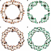
Ribbon representations of WT- and MT-FTL homopolymer crystallographic structures. A, overlay of the complete ferritin homopolymeric (24-subunit) structures of both WT- (blue) and MT-FTL (green) viewed down one of the 4-fold axes. B, overlay of the subunit structure of WT- (blue) and MT-FTL with different lengths of ordered C-terminal residues (yellow, Leu-155; red, Glu-159; green, Tyr-165), as observed crystallographically. The E-helix is missing in the mutant, and conformational differences between WT- and MT-FTL reach back to the D-helix (residue Leu-155), as detailed in the text. An additional 26 amino acids extend from Tyr-165, but were not observed in the electron density map and are not represented here. C, example of the omit σA-weighted 2FoFc electron density contoured at one standard deviation surrounding one of the 4-fold axes in the mutant ferritin structure. The WT- (blue) and MT-FTL (green) structures are represented using a ribbon diagram for better clarity to view the changes in direction of the polypeptide chain in this region. A and B were produced using SPDB-Viewer and rendered with Pov-Ray. C was produced using PyMol for Windows (34).
FIGURE 3.
FIGURE 3.
Cross-sectional representations of WT- (A and C) and MT-FTL (B and D) crystallographic structures. Each view represents a 10-Å thick slice through the middle of the 24-subunit assembly. A and B have the local 4-fold axes running horizontally and vertically and the local 2-fold axes running diagonally. C and D is a view down one of the 3-fold axes. Large but variable structural disruption is found along the 4-fold axes of the pores of the mutant but not the wild type. The C termini extended from Tyr-165 were not observed in the electron density map and are not represented here. A–D were produced using PyMol for Windows (34).
FIGURE 4.
FIGURE 4.
Iron loading-induced precipitation of MT-FTL homopolymers. Ferrous ammonium sulfate was added to WT-, TRUN-, and MT-FTL apoferritin homopolymers separately in 0.1 m Hepes, pH 7.0, and incubated for 2 h at 24 °C. Iron concentrations ranged from 0.0 to 4.0 mm with homopolymer fixed at 1 μm. Samples were centrifuged for 15 min at 10,000 × g, and soluble fractions were loaded onto native gels (3–8%) and stained with Coomassie Blue. With increasing iron concentration, MT-FTL homopolymers started to precipitate, whereas WT- and TRUN-FTL remained in solution.
FIGURE 5.
FIGURE 5.
Progression plots for iron incorporation into WT- and MT-FTL homopolymers. Ferrous ammonium sulfate was mixed with either WT- or MT-FTL apoferritins separately in 0.1 m Hepes, pH 7.0, and iron incorporation/nucleation was monitored at 24 °C by the increase in absorbance at 310 nm. Iron concentration was 1 mm, and homopolymer was 1 μm. Plots show the mean ± S.E. as error bars of triplicate experiments. Iron incorporation was substantially reduced in the mutant homopolymers. The blank monitors iron hydrolysis in the absence of protein.
FIGURE 6.
FIGURE 6.
Competition between WT- and MT-FTL homopolymers for iron incorporation. WT- and MT-FTL apoferritins alone or mixed together in equimolar amounts (WT-FTL + MT-FTL) were incubated with ferrous ammonium sulfate in 0.1 m Hepes, pH 7.0, for 30 min at 24 °C. Iron concentration was 0.1 mm, and homopolymer was 0.1 μm. After centrifugation, samples were separated by electrophoresis in native gels (3–8%). Gels were stained with Coomassie Blue (Protein Staining) and Prussian blue (Iron Staining). No iron incorporation was found for the mutant when it was in direct competition with the wild type.

References

    1. Curtis A. R., Fey C., Morris C. M., Bindoff L. A., Ince P. G., Chinnery P. F., Coulthard A., Jackson M. J., Jackson A. P., McHale D. P., Hay D., Barker W. A., Markham A. F., Bates D., Curtis A., Burn J. (2001) Nat. Genet. 28, 350–354 - PubMed
    1. Vidal R., Ghetti B., Takao M., Brefel-Courbon C., Uro-Coste E., Glazier B. S., Siani V., Benson M. D., Calvas P., Miravalle L., Rascol O., Delisle M. B. (2004) J. Neuropathol. Exp. Neurol. 63, 363–380 - PubMed
    1. Mancuso M., Davidzon G., Kurlan R. M., Tawil R., Bonilla E., Di Mauro S., Powers J. M. (2005) J. Neuropathol. Exp. Neurol. 64, 280–294 - PubMed
    1. Ohta E., Nagasaka T., Shindo K., Toma S., Nagasaka K., Ohta K., Shiozawa Z. (2008) Neurology 70, 1493–1494 - PubMed
    1. Kubota A., Hida A., Ichikawa Y., Momose Y., Goto J., Igeta Y., Hashida H., Yoshida K., Ikeda S., Kanazawa I., Tsuji S. (2009) Mov. Disord. 24, 441–445 - PubMed

Publication types

MeSH terms