. 2010 Jan 22;285(4):2545-53.
doi: 10.1074/jbc.M109.038794. Epub 2009 Nov 18.

Ikaros silences T-bet expression and interferon-gamma production during T helper 2 differentiation

Affiliations

Ikaros silences T-bet expression and interferon-gamma production during T helper 2 differentiation

Rajan M Thomas et al. J Biol Chem. .

Abstract

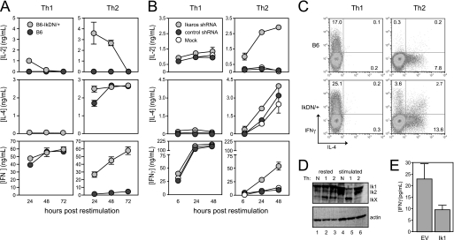
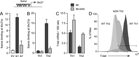
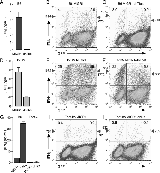
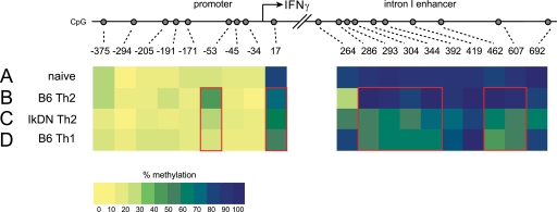
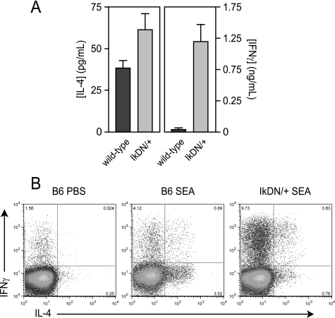
CD4+ T cells can be instructed by nonantigen-specific signals to differentiate into functionally distinct lineages with mutually exclusive patterns of cytokine production. The molecular events that drive interferon-gamma (IFN gamma) production during Th1 development are well understood, but mechanisms that silence this cytokine during Th2 polarization are not clear. In this study, we find that the tbx21 gene encoding the Th1 master regulator T-bet is a direct target of the transcriptional repressor Ikaros. In Th2 cells, which do not express T-bet, strong Ikaros binding could be detected at the endogenous tbx21 promoter, whereas this gene was not occupied by Ikaros in T-bet-expressing Th1 cells. Inhibition of Ikaros DNA binding activity during Th2 polarization resulted in loss of Ikaros promoter occupancy, increased T-bet expression, and inappropriate T-bet-dependent production of IFN gamma. Ikaros was also required for epigenetic imprinting of the ifn gamma locus during Th2 polarization, and loss of Ikaros function in vivo led to an inappropriate Th1 response to the parasite Shistosoma mansoni. These studies demonstrate that Ikaros, a factor with an established role in lymphocyte development, also regulates the development of peripheral T helper responses.


Figures

FIGURE 1.
FIGURE 1.
Ikaros DNA binding activity is required for cytokine polarization during T helper development. A, CD8-depleted spleen cells from wild-type (dark gray circles) and IkDN/+ (light gray circles) mice were stimulated with soluble anti-CD3/28 Abs with the addition of IL-12 and anti-IL-4 Ab for Th1 or IL-4, anti-IFNγ, and anti-IL-12 for Th2. B, wild-type CD8-depleted spleen cells were stimulated under either Th1 or Th2 conditions (as in A above) and transduced with retroviral vector encoding Ik1 shRNA (light gray circles), control scrambled shRNA (dark gray circles), or mock-transduced (white circles). For both A and B, cultures were harvested, washed, and restimulated with plate-bound anti-CD3/28 Ab. Secretion of IL-2, IL-4, and IFNγ secretion was measured by ELISA. C, three days following restimulation, wild-type (top panels) and IkDN/+ (bottom panels) cultures were boosted by the addition of PMA and ionomycin in the presence of monensin for 5 h, and cells were stained for surface CD4 and intracellular IL-4 (x axis) and IFNγ (y axis). Plots are gated on CD4+ cells, and the percentage of CD4+ cells that are positive for one or both cytokines is shown. D, wild-type neutral (lanes 1 and 4), Th1 (lanes 2 and 5), and Th2 (lanes 3 and 6) cells were generated as in A and rested or restimulated with plate-bound anti-CD3/28 Ab for 18 h, and Ikaros expression was assessed by immunoblotting using an Ab against the N terminus. E, purified CD4+ T cells stimulated under neutral conditions were transduced with empty MIGR1 retroviral vector (EV) or MIGR1 encoding full-length Ikaros (Ik1). Cells were rested and restimulated for 24 h with plate-bound anti-CD3/28 Ab, and IFNγ secretion was measured by ELISA. The data shown are representative of three independent experiments. Error bars are S.E. for biological replicate cultures.
FIGURE 2.
FIGURE 2.
Ikaros is a direct transcriptional repressor of the endogenous tbx21 gene. A, Ikarosnull JE131 cells were transduced with empty MIGR1 retroviral vector or reconstituted with MIGR1 encoding FLAG-tagged Ikaros lacking the DNA binding domain (Ik7) or full-length, FLAG-tagged Ikaros (Ik1). Chromatin extracts were precipitated with Ab against the FLAG epitope, and precipitated DNA was probed for the tbx21 promoter by qRT-PCR. Ikaros binding was calculated as the ratio of the specific Ab ChIP signal over the background isotype Ab control ChIP signal. Gray inverted triangles indicate putative Ikaros binding elements within the tbx21 promoter. B, wild-type (dark gray bars) and Ik7DN/+ (light gray bars) CD4+ T cells were polarized under Th1 or Th2 conditions for 24 h, and chromatin extracts were precipitated with Ab against native Ikaros. Precipitated DNA was probed for the tbx21 promoter by qRT-PCR. C and D, wild-type (dark gray bars) and Ik7DN/+ (light gray bars) Th1 and Th2 cells were restimulated on plate-bound CD3/28 Ab for 3 days. RNA was isolated, and expression of T-bet mRNA was measured by qRT-PCR (C), and T-bet protein expression in the CD4+ subset was detected by flow cytometry using fluorochrome-conjugated Ab against T-bet (D). The white histogram depicts T-bet staining of wild-type Th2 cells; the light gray histogram depicts staining of IkDN Th2 cells; and the dark gray histogram depicts wild-type Th1 cells. T-bet staining in IkDN Th1 cells was comparable with wild-type Th1 cells (not shown). The gate represents the 98% confidence interval of the isotype control stain. The data depicted are representative of two separate experiments. Error bars are S.E. for biological replicate cultures.
FIGURE 3.
FIGURE 3.
T-bet is required for IFNγ production by Ikaros mutant CD4+ T helper cells. Purified CD4+ T cells from wild-type (A–C), IkDN/+ (d–F), or tbx2−/− (G–I) mice were stimulated with PMA, ionomycin, and IL-2 for 2 days, transduced with empty MIGR1, MIGR1-dnT-bet, or MIGR1-Ik7, and cultured for an additional 3 days in IL-2. Cells were washed and restimulated with plate-bound anti-CD3/28 Ab, and secretion of IFNγ was measured 24 h later by ELISA (A, D, and G). Restimulation cultures were also boosted by the addition of PMA (3 ng/ml) and ionomycin (1 μm) in the presence of monensin (3 μm) for 5 h, and cells were assessed for surface CD4, GFP (x axis), and IFNγ (y axis) (B, C, E, F, H, and I). Plots are gated on CD4+ cells, and numbers in each upper quadrant represent the % of GFP− or GFP+ CD4+ cells that are positive for IFNγ. Numbers on the left with the triangles on the y-axes indicate the MFI of IFNγ staining in the GFP− population, and numbers on the right with triangles indicate the MFI of IFNγ staining in the GFP+ population. The data shown are representative of two separate experiments. Error bars are S.E. for biological replicate cultures.
FIGURE 4.
FIGURE 4.
Ikaros DNA binding influences DNA methylation at the ifnγ locus. Genomic DNA was purified from wild-type CD4+ naïve (A), wild-type Th2 (B), IkDN/+ Th2 (C), and wild-type Th1 (D) cells and subjected to bisulfite-mediated C > T conversion. Nested primer sets were used to amplify the ifnγ promoter and intron 1 enhancer from converted DNA, and the amplicons were cloned and sequenced. The proportion of the sequenced alleles that were methylated at each CpG site is listed in Table 1 and is depicted in pseudocolor in A–D (blue, 100% methylated; yellow, 0% methylated). The data depicted are derived from 25–27 individual cloned and sequenced alleles. Red boxes highlight regions with differential methylation.
FIGURE 5.
FIGURE 5.
Ikaros is required for Th2 polarization in vivo. Wild-type (dark gray bars) and IkDN/+ (light gray bars) mice were immunized in the footpad with soluble S. mansoni egg antigen (SEA) or phosphate-buffered saline. Six days later, draining popliteal lymph node cells were restimulated in vitro with SEA (A) or PMA/ionomycin (B), and IL-4 and IFNγ secretion was measured by ELISA at 48 h (A) or by intracellular cytokine staining at 6 h (B). Contralateral popliteal lymph node from SEA-primed mice and popliteal lymph node from phosphate-buffered saline-injected mice failed to produce detectable levels of cytokine in response to SEA stimulation in vitro (not shown). The data shown are representative of two separate experiments. Error bars are S.E. for biological replicate mice.

References

    1. Murphy K. M., Ouyang W., Farrar J. D., Yang J., Ranganath S., Asnagli H., Afkarian M., Murphy T. L. (2000) Annu. Rev. Immunol. 18, 451–494 - PubMed
    1. Afkarian M., Sedy J. R., Yang J., Jacobson N. G., Cereb N., Yang S. Y., Murphy T. L., Murphy K. M. (2002) Nat. Immunol. 3, 549–557 - PubMed
    1. Ansel K. M., Lee D. U., Rao A. (2003) Nat. Immunol. 4, 616–623 - PubMed
    1. Lee G. R., Kim S. T., Spilianakis C. G., Fields P. E., Flavell R. A. (2006) Immunity 24, 369–379 - PubMed
    1. Georgopoulos K., Bigby M., Wang J. H., Molnar A., Wu P., Winandy S., Sharpe A. (1994) Cell 79, 143–156 - PubMed

Publication types

MeSH terms