. 2010 Jan 29;285(5):3383-92.
doi: 10.1074/jbc.M109.060368. Epub 2009 Nov 18.

Helminth cysteine proteases inhibit TRIF-dependent activation of macrophages via degradation of TLR3

Affiliations

Helminth cysteine proteases inhibit TRIF-dependent activation of macrophages via degradation of TLR3

Sheila Donnelly et al. J Biol Chem. .

Abstract

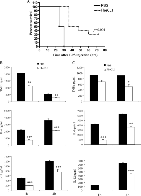
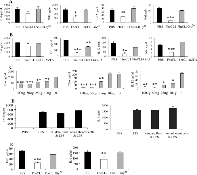
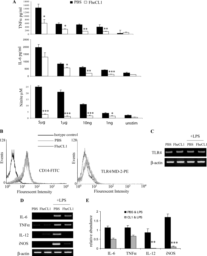
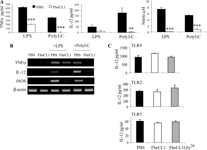
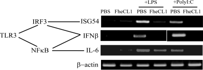
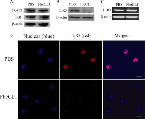
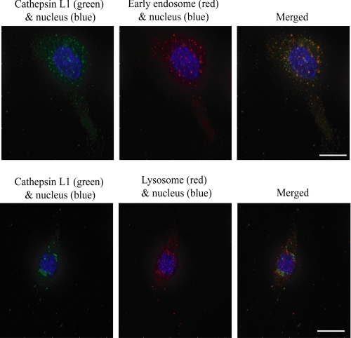
Helminth pathogens prepare a Th2 type immunological environment in their hosts to ensure their longevity. They achieve this by secreting molecules that not only actively drive type 2 responses but also suppress type 1 responses. Here, we show that the major cysteine proteases secreted from the helminth pathogens Fasciola hepatica (FheCL1) and Schistosoma mansoni (SmCB1) protect mice from the lethal effects of lipopolysaccharide by preventing the release of inflammatory mediators, nitric oxide, interleukin-6, tumor necrosis factor alpha, and interleukin-12, from macrophages. The proteases specifically block the MyD88-independent TRIF-dependent signaling pathway of Toll-like receptor (TLR)4 and TLR3. Microscopical and flow cytometric studies, however, show that alteration of macrophage function by cysteine protease is not mediated by cleavage of components of the TLR4 complex on the cell surface but occurs by degradation of TLR3 within the endosome. This is the first study to describe a parasite molecule that degrades this receptor and pinpoints a novel mechanism by which helminth parasites modulate the innate immune responses of their hosts to suppress the development of Th1 responses.


Figures

FIGURE 1.
FIGURE 1.
Helminth parasite cysteine protease protects mice from LPS-induced lethality. A, mice (n = 15) were pretreated with a single intraperitoneal injection of 50 μl of active cysteine protease enzyme (FheCL1, 1 mg/ml) or PBS 2 h prior to an intraperitoneal injection of LPS (10 mg/kg) and observed over 70 h. Data presented are representative of two independent experiments. Levels of proinflammatory mediators TNFα, IL-6, and IL-12 were measured in the sera (B) and peritoneal lavage fluid of mice (n = 6) (C) at 2 and 4 h after an intraperitoneal injection of LPS (10 mg/kg). Data are from individual mice from two independent experiments.
FIGURE 2.
FIGURE 2.
FheCL1-treated macrophages are hyporesponsive to LPS stimulation. A, peritoneal macrophages were isolated from BALB/c mice 2 h after an intraperitoneal injection of PBS, FheCL1 or an inactive mutant of cathepsin L1 (FheCL1Gly26) and stimulated ex vivo with LPS (1 μg/ml) for 12 h. The levels of secretion of IL-6, IL-12, TNFα, and nitrite into the culture supernatants were assessed by ELISA and Greiss assay, respectively. Indicated values are the means + S.E. and representative of three independent experiments. B, F. hepatica cathepsin L1 was inhibited by a 30-min incubation in the presence of cysteine protease-specific inhibitor Z-Phe-Ala-diazomethylketone (20 μm) and then injected intraperitoneally into BALB/c mice. After 2 h, peritoneal macrophages were removed and stimulated in vitro with LPS (1 μg/ml) for 12 h and secretion of proinflammatory mediators measured by ELISA and Greiss assay. Data shown are the mean ± S.E. from triplicate samples. C, BALB/c mice were given a range of concentrations of active FheCL1 (0–100 μg), and 2 h later, peritoneal macrophages were harvested and stimulated ex vivo with LPS (1 μg/ml) for 12 h. ELISA was used to measure the levels of IL-6, IL-12, and TNFα in culture supernatants. Data are presented as the mean ± S.E. of triplicate samples and are representative of three experiments. D, peritoneal macrophages were isolated from naïve BALB/c mice and incubated for 2 h in the presence of peritoneal exudate fluid or nonadherent cells extracted from the peritoneal cavity of mice given a single dose of FheCL1. The quantities of IL-6 and TNFα secreted in response to overnight stimulation with LPS (1 μg/ml) were assessed by ELISA. Data are presented as the mean ± S.E. of triplicate samples and are representative of two experiments. E, peritoneal macrophages were isolated from naïve BALB/c mice and incubated for 2 h in the presence of PBS, FheCL1, or FheCL1Gly26. The cells were subsequently stimulated with LPS (1 μg/ml), and the levels of IL-6 and TNFα in the culture media were measured by ELISA. Data are presented as the mean ± S.E. of triplicate samples and are representative of two experiments.
FIGURE 3.
FIGURE 3.
FheCL1 does not affect MyD88-dependent signaling through TLR4. A, BALB/c mice were given a single intraperitoneal injection of 50 μl of FheCL1 (1 mg/ml) or PBS and 2 h later peritoneal macrophages were removed. A, these macrophages were then stimulated with varying concentrations of LPS (0–5 μg/ml) for 12 h, and secreted cytokines were measured by ELISA. Data shown are the means + S.E. of triplicates and are representative of two individual experiments. B, macrophages removed from either PBS- or CL1-treated mice were analyzed for the surface expression of CD14 or TLR4/MD-2 by flow cytometry. C, TLR4 mRNA levels in peritoneal macrophages isolated from either PBS- or FheCL1-treated BALB/c mice incubated in the presence or absence of LPS was assessed by RT-PCR. D, peritoneal macrophages isolated from both PBS- and FheCL1-treated mice were stimulated with LPS (1 μg/ml) for 6 h, lysed, and examined for levels of mRNA expression of IL-6, TNFα, IL-12, and iNOS by RT-PCR. Data shown are representative of three independent experiments. E, densitometric analysis of amplification products for IL-6, TNFα, IL-12, and iNOS. Values for mRNA are expressed in relative absorbance units and are standardized per unit of β-actin per sample. The data are obtained from the mean of three independent experiments; FITC, fluorescein isothiocyanate; PE, phycoerythrin.
FIGURE 4.
FIGURE 4.
FheCL1 inhibits TRIF-dependent production of inflammatory mediators. A, peritoneal macrophages isolated 2 h after a single 50 μl intraperitoneal injection of either FheCL1 (1 mg/ml) or PBS were stimulated with either LPS (1 μg/ml) or poly(I·C) (25 μg/ml). B, cell supernatants were then analyzed by ELISA, and cell lysates were examined by RT-PCR for levels of mRNA expression of cytokines. C, ELISA was used to measure the quantity of IL-12 in culture media or macrophages stimulated in vitro with the TLR9 ligand CpG (1 μm), the synthetic TLR2 ligand Pam3Cys-Ser-(Lys)4(10 ng/ml) and the TLR5 ligand flagellin (10 ng/ml). Indicated values for ELISA are the means + S.E. of triplicates and are representative of three individual experiments.
FIGURE 5.
FIGURE 5.
FheCL1 treatment of macrophages inhibits TRIF-dependent signaling. Peritoneal macrophages isolated from PBS- or FheCL1-treated mice were stimulated with either LPS (1 μg/ml) or poly(I·C) (25 μg/ml) for 6 h, and expression of IFNβ, ISG54, and IL-6 mRNA were assessed by RT-PCR. These data are representative of three independent experiments.
FIGURE 6.
FIGURE 6.
FheCL1 treatment degrades TLR3 in macrophages. Cell lysates were prepared from macrophages isolated from mice, and the presence of TRIF and TRAF3 protein was determined by Western blot (A). B, detection of TLR3 protein in peritoneal macrophages was examined by Western blot, and expression of mRNA for TLR3 was assessed by RT-PCR. C, peritoneal macrophages isolated from PBS-treated (upper panels) or FheCL1-treated (lower panels) mice were immunostained for the presence of TLR3 (red). Cells were also stained with 4′,6-diamidino-2-phenylindole (blue) to detect nuclei and imaged using a Nikon A1 confocal scanning laser microscope. Scale bar is 10 μm. These data are representative of two independent experiments.
FIGURE 7.
FIGURE 7.
FheCL1 is targeted to the endolysosomal compartment of macrophages. Peritoneal macrophages were isolated from BALB/c mice 2 h after an intraperitoneal injection of FheCL1. After fixation and permeabilization, cells were stained for His6 tag (FheCL1, green), EEA-1 (early endosomes, red, upper panel) and mannose-6-phosphate receptor (lysosomes, red, lower panel). Cells were also stained with 4′,6-diamidino-2-phenylindole (blue) to detect nuclei and imaged using a DeltaVision personal DV microscope. Scale bar is 10 μm.

References

    1. Balic A., Harcus Y. M., Taylor M. D., Brombacher F., Maizels R. M. (2006) Int. Immunol. 18, 1421–1431 - PubMed
    1. Carvalho L., Sun J., Kane C., Marshall F., Krawczyk C., Pearce E. J. (2009) Immunol. 126, 28–34 - PMC - PubMed
    1. Siracusa M. C., Reece J. J., Urban J. F., Jr., Scott A. L. (2008) J. Leukoc. Biol. 84, 1422–1433 - PMC - PubMed
    1. Kuroda E., Yoshida Y., En Shan B., Yamashita U. (2001) Parasite Immunol. 23, 305–311 - PubMed
    1. Goodridge H. S., Marshall F. A., Else K. J., Houston K. M., Egan C., Al-Riyami L., Liew F. Y., Harnett W., Harnett M. M. (2005) J. Immunol. 174, 284–293 - PubMed

Publication types