. 2010 Jan;103(1):419-23.
doi: 10.1152/jn.00718.2009. Epub 2009 Nov 18.

Vesicle pool size at the salamander cone ribbon synapse

Affiliations

Vesicle pool size at the salamander cone ribbon synapse

Theodore M Bartoletti et al. J Neurophysiol. 2010 Jan.

Abstract

Cone light responses are transmitted to postsynaptic neurons by changes in the rate of synaptic vesicle release. Vesicle pool size at the cone synapse constrains the amount of release and can thus shape contrast detection. We measured the number of vesicles in the rapidly releasable and reserve pools at cone ribbon synapses by performing simultaneous whole cell recording from cones and horizontal or off bipolar cells in the salamander retinal slice preparation. We found that properties of spontaneously occurring miniature excitatory postsynaptic currents (mEPSCs) are representative of mEPSCs evoked by depolarizing presynaptic stimulation. Strong, brief depolarization of the cone stimulated release of the entire rapidly releasable pool (RRP) of vesicles. Comparing charge transfer of the EPSC with mEPSC charge transfer, we determined that the fast component of the EPSC reflects release of approximately 40 vesicles. Comparing EPSCs with simultaneous presynaptic capacitance measurements, we found that horizontal cell EPSCs constitute 14% of the total number of vesicles released from a cone terminal. Using a fluorescent ribeye-binding peptide, we counted approximately 13 ribbons per cone. Together, these results suggest each cone contacts a single horizontal cell at approximately 2 ribbons. The size of discrete components in the EPSC amplitude histogram also suggested approximately 2 ribbon contacts per cell pair. We therefore conclude there are approximately 20 vesicles per ribbon in the RRP, similar to the number of vesicles contacting the plasma membrane at the ribbon base. EPSCs evoked by lengthy depolarization suggest a reserve pool of approximately 90 vesicles per ribbon, similar to the number of additional docking sites further up the ribbon.


Figures

Fig. 1.
Fig. 1.
Spontaneous miniature excitatory postsynaptic currents (mEPSCs) are indistinguishable from mEPSCs evoked by depolarization of a voltage-clamped cone. A: spontaneously occurring mEPSCs recorded from a horizontal cell in bright light. B: mEPSCs evoked in the same horizontal cell by depolarizing a presynaptic cone to −30 mV in the presence of a bright background light. C: there was no significant difference between the cumulative amplitude histograms of spontaneously occurring mEPSCs (—) and mEPSCs recorded during the depolarizing test step (- - -). Cumulative amplitude histograms in this figure were averaged from 6 cone/horizontal cell pairs. D: quantal amplitudes determined from the steep initial slope of the rise in the relationship between variance and mean amplitude of horizontal cell EPSCs evoked by pairs of test steps to −40, −30, or −20 mV applied to cones (8.2 ± 2.2 responses at each potential, 30-s interstimulus interval, n = 6 cone/horizontal cell pairs).
Fig. 2.
Fig. 2.
Fully activating cone ICa evokes release with probability (P) ∼1. A: representative postsynaptic recording in a horizontal cell in response to a train of pulses to −10 mV from −70 mV applied to a voltage-clamped presynaptic cone. The pulses were 25 ms in duration and 50 ms apart. B: cumulative amplitude of EPSCs evoked during pulse trains were plotted against time, and a straight line was fit to the final 3 data points when replenishment balances release. The y intercept of this line shows the size of the vesicle pool available for release at the beginning of the pulse train. The 1st pulse of the stimulus train released nearly all the available vesicles. C: elevating [Ca2+]e to 5 mM enhanced cone ICa (bottom) but did not increase the amplitude of the EPSC recorded simultaneously in a horizontal cell (HC). This indicates that all available vesicles were released by depolarizing pulses from −70 to −10 mV in normal Ca2+ levels. Control: black traces. 5 mM Ca2+: gray traces. Passive Rm and Cm were subtracted from the cone membrane current using a P/8 leak subtraction protocol.
Fig. 3.
Fig. 3.
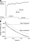
The EPSC consists of a fast component due to release of the rapidly releasable pool (RRP) followed by a slower release component. We integrated the depolarization-evoked horizontal cell EPSC (A) to measure charge transfer. To separate fast and slow release components, we fit EPSC charge transfer with a sum of 2 exponentials (B). — in B shows the biexponential fit to the EPSC. We also illustrate the 2 components of this biexponential function separately: - - -, the fast exponential (702 pC, τ = 3.4 ms); ···, the slower component (4,189 pC, τ = 121 ms).
Fig. 4.
Fig. 4.
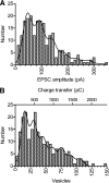
EPSC amplitude histograms show multiple peaks consistent with the presence of multiple ribbon contacts between individual cones and horizontal cells. A: the amplitude distribution of EPSCs fit using a multiple Gaussian function (—, R2 = 0.93, n = 217) with individual Gaussian components averaging 46.3 ± 17.8 (SD) pA. B: the histogram of charge transfer measurements of the initial fast component of the EPSC was also fit with a multiple Gaussian function (—, R2 = 0.90, n = 217). The amplitude of the fast component was determined from the fit to charge transfer using a double exponential function as described in Fig. 3. After scaling charge transfer measurements by 16 pC/vesicle, individual Gaussian components averaged 15.2 ± 5.7 (SD) vesicle.
Fig. 5.
Fig. 5.
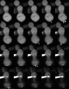
Example of cone ribbons labeled by HyLite488 conjugated to a peptide that binds to the ribbon protein, ribeye. The ribeye-binding peptide was introduced through the patch pipette (the large bright structure at the left in the bottom 2 rows of images). The figure shows 20 confocal slices from a single cone obtained at 0.5-μM increments. Top left: the uppermost slice. Ribbons extended through multiple sections and were distinguished in 3 dimensions. The appearance of 14 different ribbons is shown by small arrows at the base of the cone. Images were acquired using 488-nm excitation and 525-nm emission with image durations of 2 s per confocal slice. Scale bar = 5 μm.
Fig. 6.
Fig. 6.
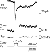
Comparison of the amount of release measured from a single horizontal cell (HC) EPSC to the total amount of release from a cone measured using capacitance techniques. Top: the EPSC evoked in a horizontal cell by a depolarizing test step applied to the cone (−70 to −10 mV, 25 ms). The depolarizing step stimulated a simultaneous increase in cone Cm (2nd trace) but no appreciable change in cone Rser (3rd trace).
Fig. 7.
Fig. 7.
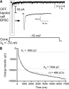
Measurement of the reserve pool. A: EPSC evoked in an off bipolar cell by prolonged depolarizing test steps (7 s, −70 to −10 mV) applied to the cone. Inset: the initial fast component of the EPSC. B: charge transfer during the EPSC fit with a sum of 2 exponentials plus a straight line. We fit the 1st 200 ms with a double-exponential function to measure the RRP. In this example, the fast component was 1860 pC in amplitude (A1) with a time constant of 6.4 ms. We then fixed the best-fit parameters for the fast exponential and fit the entire 7-s trial with a double-exponential plus straight line function. In this example, the slower component had a time constant of 653 ms and was 7,656 pC (A2), fourfold larger than the fast component. The straight line, which likely reflects the rate of replenishment, had a slope (m) of −498 pC/s.

Similar articles

Cited by

References

    1. Ahnelt P, Kolb H. Horizontal cells and cone photoreceptors in primate retina: a Golgi-electron microscope study of spectral connectivity. J Comp Neurol 343: 406–427, 1994 - PubMed
    1. Brandt A, Khimich D, Moser T. Few CaV1.3 channels regulate the exocytosis of a synaptic vesicle at the hair cell ribbon synapse. J Neurosci 25: 11577–11585, 2005 - PMC - PubMed
    1. Cadetti L, Bartoletti TM, Thoreson WB. Quantal mEPSCs and residual glutamate: how horizontal cell responses are shaped at the photoreceptor ribbon synapse. Eur J Neurosci 27: 2575–2586, 2008 - PMC - PubMed
    1. Cadetti L, Tranchina D, Thoreson WB. A comparison of release kinetics and glutamate receptor properties in shaping rod-cone differences in EPSC kinetics in the salamander retina. J Physiol 569: 773–788, 2005 - PMC - PubMed
    1. Church PJ, Stanley EF. Single L-type calcium channel conductance with physiological levels of calcium in chick ciliary ganglion neurons. J Physiol 496: 59–68, 1996 - PMC - PubMed

Publication types

LinkOut - more resources