Comparative Study
. 2009 Nov 18;29(46):14534-44.
doi: 10.1523/JNEUROSCI.1546-09.2009.

APP anterograde transport requires Rab3A GTPase activity for assembly of the transport vesicle

Affiliations
Comparative Study

APP anterograde transport requires Rab3A GTPase activity for assembly of the transport vesicle

Anita Szodorai et al. J Neurosci. .

Abstract

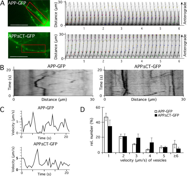
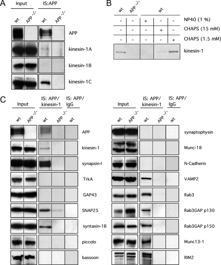
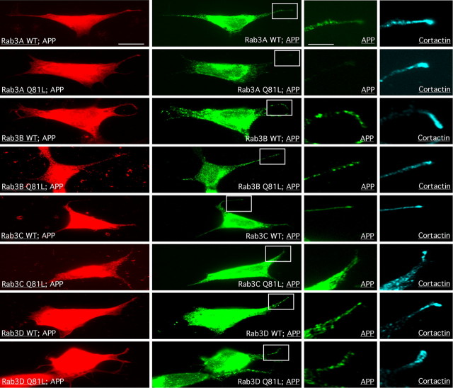
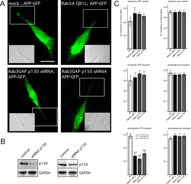
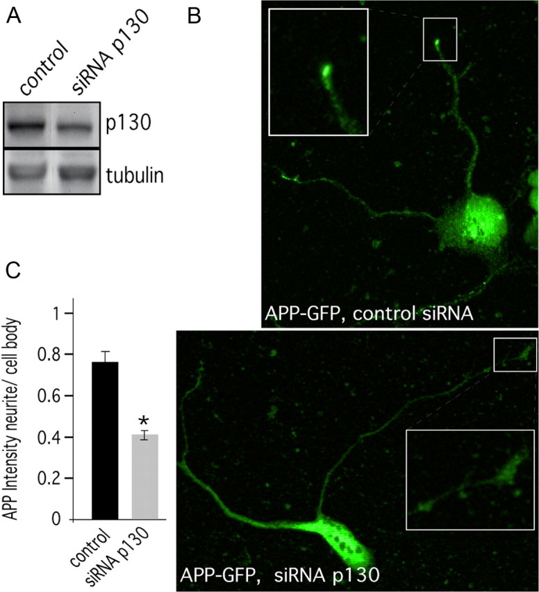
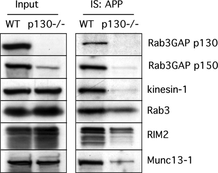
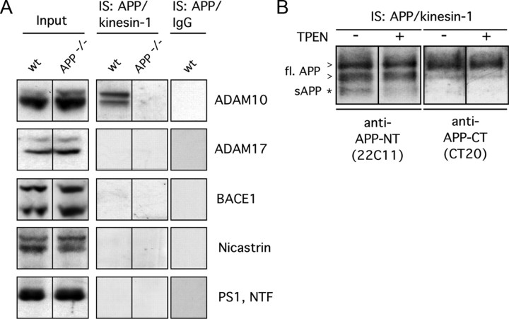
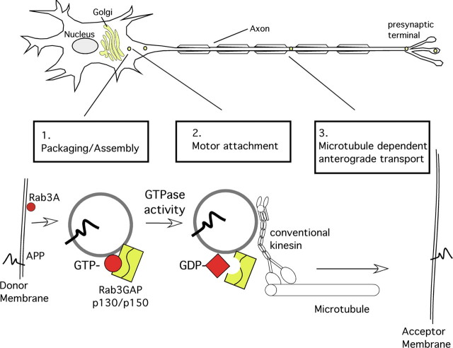
The amyloid precursor protein (APP) is anterogradely transported by conventional kinesin in a distinct transport vesicle, but both the biochemical composition of such a vesicle and the specific kinesin-1 motor responsible for transport are poorly defined. APP may be sequentially cleaved by beta- and gamma-secretases leading to accumulation of beta-amyloid (Abeta) peptides in brains of Alzheimer's disease patients, whereas cleavage of APP by alpha-secretases prevents Abeta generation. Here, we demonstrate by time-lapse analysis and immunoisolations that APP is a cargo of a vesicle containing the kinesin heavy chain isoform kinesin-1C, the small GTPase Rab3A, and a specific subset of presynaptic protein components. Moreover, we report that assembly of kinesin-1C and APP in this vesicle type requires Rab3A GTPase activity. Finally, we show cleavage of APP in transport vesicles by alpha-secretase activity, likely mediated by ADAM10. Together, these data indicate that maturation of APP transport vesicles, including recruitment of conventional kinesin, requires Rab3 GTPase activity.


Figures

Figure 1.
Figure 1.
APP can be transported by the fast axonal transport machinery in the absence of its intracellular C terminus. Mouse primary cortical neurons (DIV7) expressing APP–GFP and/or APPΔCT–GFP were analyzed by time-lapse microscopy 18 h after transfection. A, Fluorescence micrographs of APP–GFP- and APPΔCT-GFP-expressing neurons. For tracking and velocity analyses, a region of interest (red box) was selected. Sequential images of the region of interest are shown to the right. Colored arrowheads indicate the single vesicles that have been examined. Time interval between images was 200 ms, 5 images/s. B, Representative kymographs showing APP–GFP and APPΔCT-GFP movement. C, Velocities of APP–GFP- and APPΔCT-GFP-containing vesicles, assayed over a period of 22 s. D, Histogram showing quantification of the number of recorded vesicles moving at velocities of 1, 2, 3, 4, 5, or >6 μm/s. No statistical significant differences between the velocities of APP–GFP- and APPΔCT-containing transport vesicles could be determined (Student's t test, p ≥ 0.2). Scale bar, 25 μm.
Figure 2.
Figure 2.
APP is a cargo of an axonal transport vesicle containing presynaptic components and is associated with kinesin-1C. A, Immunoisolations (IS) of APP using an anti-APP antibody (CT20) from wild-type (wt) and APP knock-out (APP−/−) mouse brain homogenates. Each lysate (180) was loaded as input control (Input). Western blot analyses of APP immunoisolates with antibodies against specific kinesin-1s revealed that APP is mainly associated with kinesin-1C. B, Immunoisolated APP containing membrane preparations from wild-type mice (wt) were treated with 1% (v/v) NP-40 (lane 3), 15 mm CHAPS (lane 4), 1.5 mm CHAPS (below the critical micellar concentration) (lane 5), or no detergent (lane 1). An immmunoisolate using anti-APP antibody obtained from APP knock-out mice (APP−/−) is shown as a control (lane 2). C, Total mouse brain membrane fractions were separated on a linear iodixanol gradient (supplemental Fig. 2, available at www.jneurosci.org as supplemental material), and low-density membrane fractions (fractions 1–5) were pooled and used for a two-step immunoisolation: vesicles were first immunoisolated with an anti-APP antibody (IS:APP). After these, immunoisolated membranes were eluted and subject to immunoisolation with an anti-kinesin-1 antibody (H2) (IS: APP/kinesin-1) (for details, see Materials and Methods). Coimmunoisolated proteins were separated by SDS-PAGE and subjected to Western blot analysis. Brain homogenates from APP knock-out (APP−/−) mice were used for immunoisolation with anti-APP antibody as a control for nonspecific binding. To test for specificity of anti-kinesin-1 immunoisolations, a non-immune IgG fraction (IgG) was used (IS:APP/IgG).
Figure 3.
Figure 3.
Rab3-subtype-specific influence on APP localization. To determine specific Rab3 family members that might be essential for anterograde transport of APP, we tested the influence of both wild-type (wt) and GTPase-deficient mutant (Q81L) versions of GFP-tagged Rab3A–Rab3D on APP-HA localization at the distal end of growing neurites. Neuroblastoma cells (SH-SY5Y) were cotransfected with the different Rab3 constructs together with APP-HA cDNA and analyzed by immunocytochemistry using anti-GFP (red), anti-HA (green), and as a growth cone marker anti-cortactin (cyan) antibodies. Notably, only transfection with the Rab3A Q81L construct caused a reduction in the level of APP that normally accumulates at the distal ends of neurites. To visualize the neurites more clearly, the regions of interest (boxed) are shown at higher magnification in the two right columns. Scale bar, 10 μm. Higher-magnification bar, 3.3 μm.
Figure 4.
Figure 4.
Rab3 GTPase activity is essential for APP fast anterograde transport. Neuroblastoma cells (SH-SY5Y) were cotransfected with APP–GFP cDNA and empty vector (mock), Rab3A Q81L cDNA, shRNA constructs directed against Rab3GAP p130, or Rab3GAP p150. A, Mock-transfected cells expressing APP–GFP displayed an accumulation of APP–GFP at the tips of neurites (box), whereas inhibition of Rab3A GTPase activity by expression of mutant Rab3A Q81L or silencing of Rab3GAP p130 or p150 caused a decrease of APP–GFP levels at the tips. Differential interference contrast images of the regions of interest/of the indicated regions (boxed) are shown as insets. Scale bar, 10 μm. B, The silencing efficiency of the human Rab3GAP p130 or Rab3GAP p150 shRNA was of ∼40–60%. Taking into account that the transfection rate with the vector-based shRNA constructs was 50–60%, the knockdown efficiency per transfected cell was estimated to be ∼70–80%. GAPDH, Glyceraldehyde-3-phosphate dehydrogenase. C, APP–GFP (left) or synaptophysin–GFP (right) fusion proteins were analyzed by time-lapse microscopy (5 frames/s) in mock-transfected neuroblastoma cells (white columns), cells coexpressing Rab3A Q81L (black columns), or cells with knockdown [RNA interference (RNAi)] of Rab3GAP p130 (dark gray columns) or Rab3GAP p150 (light gray columns). Kymographs from single cells (n = 12) were analyzed. The relative frequencies of stationary, retrogradely, or anterogradely transported APP–GFP (left) or synaptophysin–GFP (right) containing vesicles was determined. The range of velocities for synaptophysin–GFP (syn) is lower (1–5 μm/s) than the range seen with APP (1–10 μm/s). Therefore, we determined only the relative frequency of anterograde APP–GFP vesicles moving at velocities ≥4 μm. Error bars represent SEM. ***p < 0.001, t test.
Figure 5.
Figure 5.
Inhibition of Rab3 GTPase activity reduces the levels of APP at the tips of neurites. Mouse primary cortical neurons (DIV1) were transfected with siRNA directed against Rab3GAP p130 or nonfunctional control siRNA. A, Western blot analysis indicated that the silencing effect of mouse Rab3GAP p130 was ∼35%. B, Immunocytochemistry of the siRNA transfected stage 1 primary neurons using antibody directed against the APP C terminus (CT20) reveals that the cells treated with Rab3GAP p130 siRNA have less APP accumulation at the tips of neurites. For clarity, the tips of the neurites (boxed) are shown at a higher magnification as inset. C, For quantification, the intensity of APP in the cell body and in the tip of the neurites was measured using NIH ImageJ. The ratio of APP intensity at tips/cell bodies was reduced ∼40% in cells treated with Rab3GAP p130 siRNA compared with control siRNA transfected cells. Error bars represent SEM. *p > 0.01, t test. Scale bar, 5 μm.
Figure 6.
Figure 6.
Loss of kinesin-1 from APP transport vesicles in Rab3GAP p130 knock-out mice. Low-density membrane fractions from wild-type (wt) or Rab3GAP p130 knock-out mouse (p130−/−) brain homogenates were immunoisolated with anti-APP antibodies (IS:APP) and subjected to Western blot analyses using antibodies directed against Rab3GAP p130, Rab3GAP p150, kinesin-1, Rab3, and two putative cargoes of APP transport vesicles, RIM2 and Munc13-1 (see Fig. 1). Total lysate (180) from each brain was loaded in the lanes of the left panel as input control (Input).
Figure 7.
Figure 7.
APP and ADAM10 are cargoes of a common transport vesicle. A, Western blot analyses of APP/kinesin-1 double immunoisolates (IS:APP/kinesin-1) with anti-ADAM10, anti-ADAM17, anti-BACE1, anti-PS1, and anti-Nicastrin antibodies. As control for APP/kinesin-1 double immunoisolations, brain homogenates from APP knock-out mice (APP−/−) and non-immune IgGs (IS:IgG) were used. Each lysate (180) was loaded as input control (Input). wt, Wild type. B, Western blot analyses of APP/kinesin-1 double-immunoisolated membrane preparations (IS:APP/kinesin-1) with an antibody directed against the APP N terminus (22C11), recognizing full-length (fl.) APP (arrowhead) and cleaved sAPP (asterisk), as well as an antibody directed against the APP C terminus, recognizing full-length APP (CT20) but not sAPP. TPEN, a zinc chelator blocking α-secretase activity, was added directly after brain homogenization at a concentration of 10 μm when indicated (+).
Figure 8.
Figure 8.
Model of APP anterograde transport vesicle assembly. Data presented here suggest that, in neurons, APP is sorted into an FAT vesicle containing Rab3A and other presynaptic components. 1, In the first step (likely in the late Golgi apparatus), the GTP-bound form of Rab3A (red circle) mediates packaging and assembly of APP and a specific set of presynaptic components (i.e., syntaxin-1, SNAP25, synapsin-1, Munc13-1, and RIM2) in a common vesicle. 2, After this, Rab3A-GTP hydrolysis catalyzed by dimerized Rab3GAP p130 and p150 (yellow rectangle) causes a conformational change of Rab3A-GTP (red circle) to Rab3A-GDP (red square), accompanied by recruitment of a specific kinesin-1 isoform variant (kinesin-1C) to the APP transport vesicle. 3, Finally, the assembled FAT vesicle is transported by this conventional kinesin via microtubule tracks to its target membrane at the presynaptic terminal, in which the APP transport vesicle fuses with the presynaptic acceptor membrane.

Similar articles

Cited by

References

    1. Ali BR, Seabra MC. Targeting of Rab GTPases to cellular membranes. Biochem Soc Trans. 2005;33:652–656. - PubMed
    1. Amaratunga A, Morin PJ, Kosik KS, Fine RE. Inhibition of kinesin synthesis and rapid anterograde axonal transport in vivo by an antisense oligonucleotide. J Biol Chem. 1993;268:17427–17430. - PubMed
    1. Back S, Haas P, Tschäpe JA, Gruebl T, Kirsch J, Müller U, Beyreuther K, Kins S. beta-amyloid precursor protein can be transported independent of any sorting signal to the axonal and dendritic compartment. J Neurosci Res. 2007;85:2580–2590. - PubMed
    1. Bloom GS, Richards BW, Leopold PL, Ritchey DM, Brady ST. GTP gamma S inhibits organelle transport along axonal microtubules. J Cell Biol. 1993;120:467–476. - PMC - PubMed
    1. Burstein ES, Brondyk WH, Macara IG, Kaibuchi K, Takai Y. Regulation of the GTPase cycle of the neuronally expressed Ras-like GTP-binding protein Rab3A. J Biol Chem. 1993;268:22247–22250. - PubMed

Publication types

MeSH terms