. 2009 Dec 15;183(12):7787-98.
doi: 10.4049/jimmunol.0902318.

Mesenchymal stem cells derived from human gingiva are capable of immunomodulatory functions and ameliorate inflammation-related tissue destruction in experimental colitis

Affiliations

Mesenchymal stem cells derived from human gingiva are capable of immunomodulatory functions and ameliorate inflammation-related tissue destruction in experimental colitis

Qunzhou Zhang et al. J Immunol. .

Erratum in

  • J Immunol. 2010 Feb 1;184(3):1656

Abstract

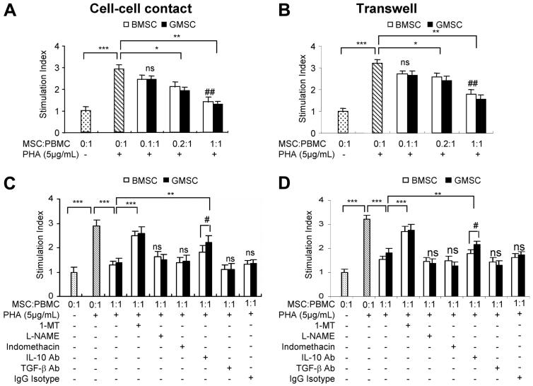
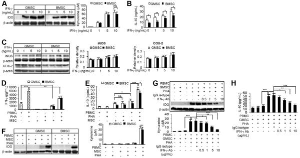
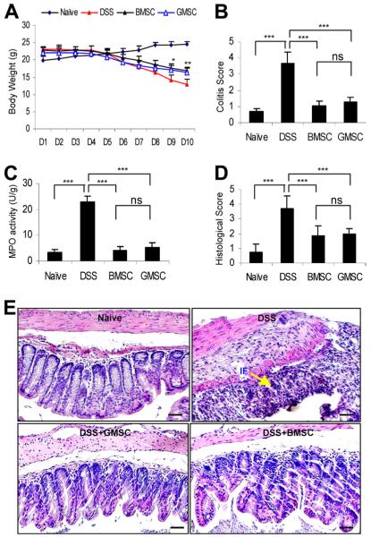
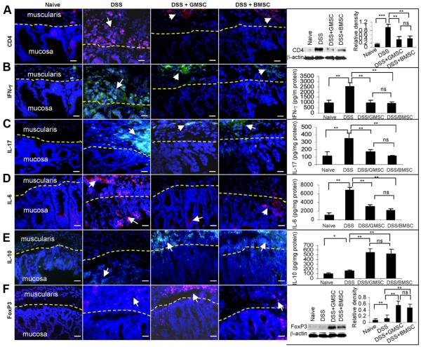
Aside from the well-established self-renewal and multipotent differentiation properties, mesenchymal stem cells exhibit both immunomodulatory and anti-inflammatory roles in several experimental autoimmune and inflammatory diseases. In this study, we isolated a new population of stem cells from human gingiva, a tissue source easily accessible from the oral cavity, namely, gingiva-derived mesenchymal stem cells (GMSCs), which exhibited clonogenicity, self-renewal, and multipotent differentiation capacities. Most importantly, GMSCs were capable of immunomodulatory functions, specifically suppressed peripheral blood lymphocyte proliferation, induced expression of a wide panel of immunosuppressive factors including IL-10, IDO, inducible NO synthase (iNOS), and cyclooxygenase 2 (COX-2) in response to the inflammatory cytokine, IFN-gamma. Cell-based therapy using systemic infusion of GMSCs in experimental colitis significantly ameliorated both clinical and histopathological severity of the colonic inflammation, restored the injured gastrointestinal mucosal tissues, reversed diarrhea and weight loss, and suppressed the overall disease activity in mice. The therapeutic effect of GMSCs was mediated, in part, by the suppression of inflammatory infiltrates and inflammatory cytokines/mediators and the increased infiltration of regulatory T cells and the expression of anti-inflammatory cytokine IL-10 at the colonic sites. Taken together, GMSCs can function as an immunomodulatory and anti-inflammatory component of the immune system in vivo and is a promising cell source for cell-based treatment in experimental inflammatory diseases.


Figures

FIGURE 1
FIGURE 1
Expression of stem cell markers in human gingival tissues. A, H&E staining of paraffin sections of human gingival tissues. MBV, Microvascular blood vessel; BV, blood vessel. B, Frozen sections were immunostained with mouse mAbs specific for human Oct-4, SSEA-4, and Stro-1 or an isotype-matched mouse IgG, followed by incubation with FITC-conjugated secondary Abs. Images were observed under a fluorescence microscope. Scale bar, 100 μm. The results are representative of at least five independent experiments.
FIGURE 2
FIGURE 2
Isolation and subcloning of MSCs from human gingival tissues. A, Subcloning and culture of MSCs from gingival tissues in α-MEM supplemented with 10% FBS, 1× nonessential amino acids and antibiotics. Scale bar, 100 μm. B, Capability of colony formation of gingiva-derived cells. C, Population doublings of GMSCs. D, Expression of stem cell markers in GMSCs. Cells cultured in an 8-well slide chamber were fixed and immunostained with specific Abs for human Stro-1, SSEA-4, Oct-4, or hTERT. Cells were incubated with rhodamine- or FITC-conjugated secondary Abs and then observed under a fluorescence microscope. Scale bar, 100 μm. E, Semiquantification of positive signals in at least five random high-power fields and expressed as the percentage of total DAPI-positive cells (mean ± SD). F, Expression of cell surface markers on GMSCs as determined by flow cytometry. G, Quantification of percentage of cells expressing respective surface markers from independent experiments from flow cytometry data (mean ± SD). The results are representative of at least five independent experiments.
FIGURE 3
FIGURE 3
Multipotent differentiation of GMSCs. A, Adipogenic differentiation of GMSCs. After culture under normal growth conditions (control, lanes 1 and 3) or adipogenic differentiation (lanes 2 and 4) conditions for 2 wk, adipocyte differentiation was determined by Oil Red O staining and RT-PCR analysis of specific genes. The graph shows the quantification of the Oil Red O dye content in differentiated adipocytes from independent experiments (mean ± SD). B, Osteogenic differentiation of GMSCs. After culture under normal growth conditions (control, lanes 1 and 3) or osteogenic differentiation conditions (lanes 2 and 4) for 4–5 wk, osteogenic differentiation was determined by Alizarin Red S staining and RT-PCR analysis of specific genes. The graph shows the quantification of the Alizarin Red S dye content in differentiated osteocytes from independent experiments (mean ± SD). OCN, Osteocalcin. Scale bar, 50 μm. C, Endothelial differentiation of GMSCs after culture in endothelial cell culture conditions for 7 days. Cells were immunostained with a mouse monoclonal IgG for human CD31, followed by incubation with FITC-conjugated secondary Ab, and then observed under a fluorescence microscope. Scale bar, 100 μm. D, Neural differentiation of GMSCs after culture in neural cell culture conditions for 14 days. Cells were immunostained with different primary Abs for neural markers, including GFAP, neurofilament M (NF-M), and β-tubulin III, followed by incubation with rhodamine- or FITC-conjugated secondary Abs and then observed under a fluorescence microscope. Scale bar, 100 μm. E, In vivo transplantation of GMSCs. Approximately 2.0 × 10⁶ stem cells mixed with 40 mg of HA/TCP ceramic powder were s.c. transplanted into the dorsal surface of 8- to 10-wk-old female immunocompromised mice. Four weeks later, the transplants were harvested and cells were recovered for secondary transplantation. H&E staining was performed for histological examination. The cells of human origin were confirmed by immunostaining with a specific Ab for human mitochondria. Scale bar, 50 μm. F, Immunohistochemical studies of the expression of human type I collagen and Oct-4 in GMSC-derived transplants. The results are representative of at least three independent experiments.
FIGURE 4
FIGURE 4
Inhibitory effects of GMSCs on PHA-stimulated PBMC proliferation. A and B, 2 × 10⁵ PBMCs were cultured alone or cocultured with increasing numbers of GMSCs or BMSCs under both cell-cell contact (A) and Transwell (B) conditions in the presence or absence of 5 μg/ml PHA for 72 h. Afterward, cell numbers were counted using a Cell Counting Kit-8. *, p < 0.05; **, p < 0.01; ***, p < 0.001; ns, no significant difference; ##, p < 0.05 compared with Transwell. C and D, GMSCs or BMSCs were pretreated for 2 h wit h 1-MT (1 mM), l-NAME (500 μM), indomethacin (10 μM), or neutralizing Abs for IL-10 or TGF-β1 (10 μg/ml), followed by coculturing with the same number of PBMCs (1/1) under both cell-cell contact (C) and Transwell (D) conditions in the presence or absence of 5 μg/ml PHA for 72 h. Afterward, cell numbers were counted using a Cell Counting Kit-8. *, p < 0.05; **, p < 0.01; ***, p < 0.001; #, p < 0.05; ns, no significant difference (mean ± SD). The results are representative of at least three independent experiments.
FIGURE 5
FIGURE 5
IFN-γ-induced IDO expression and IL-10 secretion by GMSCs. A–C, GMSCs or BMSCs were stimulated with increasing concentrations of IFN-γ for 24 h. Then the expression of IDO protein was determined by Western blot, while the IDO activity was analyzed by measuring the concentration of kynurenine in the conditioned medium (A). IFN-γ-induced IL-10 secretion in the supernatants was determined by using ELISA (B), whereas the expression of iNOS and COX-2 in MSCs in response to IFN-γ was determined by Western blot (C). D–F, Two × 105 PBMCs were cultured alone or cocultured with the same number of GMSCs or BMSCs under cell-cell contact conditions in the presence or absence of 5 μg/ml PHA for 72 h. Afterward, the concentrations of IFN-γ (D) and IL-10 (E) in the supernatants were determined by using ELISA, whereas IDO protein expression and activity were determined by Western blot and kynurenine assay, respectively (F). G and H, PBMCs were pretreated for 2 h with different concentrations of specific neutralizing Ab for human IFN-γ (0.5–10 μg/ml) or an isotype-matched mouse IgG (10 μg/ml), followed by coculturing with the same number of GMSCs (1/1) under cell-cell contact conditions in the presence or absence of 5 μg/ml PHA for 72 h. Then IDO protein expression and activity were determined by Western blot and kynurenine assay, respectively (G), whereas the concentration of IL-10 in the supernatants was determined by using ELISA (H). *, p < 0.05; **, p < 0.01; ***, p < 0.001; ns, no significant difference (mean ± SD). The results are representative of at least three independent experiments.
FIGURE 6
FIGURE 6
Treatment with GMSCs ameliorates DSS-induced experimental colitis in C57BL/6 mice. Colitis was induced by oral administration of 3% DSS in drinking water for 7 days. Two × 106 of GMSCs or BMSCs in 200 μl of PBS were i.p. injected into mice 1 day after initiation of DSS treatment. Mice without any treatment (naive mice) or mice that received 200 μl of PBS served as controls. At day 10, mice were sacrificed. A and B, Clinical progression of the disease was monitored by body weight changes (A) and colitis score evaluation (B), whereas in A, *, p < 0.05 and **, p < 0.01 compared with DSS alone. C, Colonic MPO activity assays. D and E, Histopathological analysis of colitis. IF, Inflammation. Scale bar, 200 μm. *, p < 0.05; **, p < 0.01; ***, p < 0.001; ns, no significant difference (mean ± SD). The results are representative of at least three independent experiments.
FIGURE 7
FIGURE 7
GMSC treatment attenuates colonic inflammatory responses but induces Treg responses in DSS-induced experimental colitis in C57BL/6 mice. Colitis was induced by oral administration of 3% DSS in drinking water for 7 days. Two × 106 of GMSCs or BMSCs in 200 μl of PBS were i.p. injected into mice 1 day after initiation of DSS treatment. Mice without any treatment (naive mice) or mice that received 200 μl of PBS served as controls. At day 10, mice were sacrificed. A, Immunofluorescence staining and Western blot analysis of the infiltrated CD4+ T lymphocytes in inflamed colons. B–E, Immunofluorescence staining and ELISA of IFN-γ, IL-17, IL-6, and IL-10 in inflamed colons. F, Immunofluorescence staining and Western blot analysis of FoxP3 in inflamed colon tissues. Scale bars, 100 μm. *, p < 0.05; **, p < 0.01; ***, p < 0.001; ns, no significant difference (means ± SD). The results are representative of at least three independent experiments.

References

    1. Prockop DJ. Marrow stromal cells as stem cells for nonhematopoietic tissues. Science. 1997;276:71–74. - PubMed
    1. Pittenger MF, Mackay AM, Beck SC, Jaiswal RK, Douglas R, Mosca JD, Moorman MA, Simonetti DW, Craig S, Marshak DR. Multilineage potential of adult human mesenchymal stem cells. Science. 1999;284:143–147. - PubMed
    1. Friedenstein AJ, Chailakhjan RK, Lalykina KS. The development of fibroblast colonies in monolayer cultures of guinea-pig bone marrow and spleen cells. Cell Tissue Kinet. 1970;3:393–403. - PubMed
    1. Fernandes KJ, McKenzie IA, Mill P, Smith KM, Akhavan M, Barnabé-Heider F, Biernaskie J, Junek A, Kobayashi NR, Toma JG, et al. A dermal niche for multipotent adult skin-derived precursor cells. Nat. Cell Biol. 2004;6:1082–1093. - PubMed
    1. Toma JG, McKenzie IA, Bagli D, Miller FD. Isolation and characterization of multipotent skin-derived precursors from human skin. Stem Cells. 2005;23:727–737. - PubMed

Publication types

MeSH terms

Substances