. 2009 Dec 15;183(12):7635-8.
doi: 10.4049/jimmunol.0804251.

Cutting edge: normal regional lymph node enrichment of antigen-specific regulatory T cells with autoimmune disease-suppressive capacity

Affiliations

Cutting edge: normal regional lymph node enrichment of antigen-specific regulatory T cells with autoimmune disease-suppressive capacity

Karen M Wheeler et al. J Immunol. .

Abstract

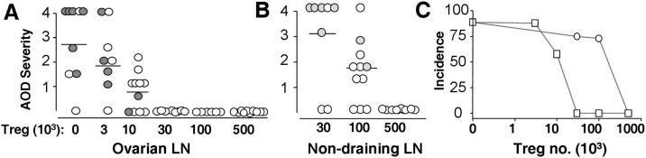
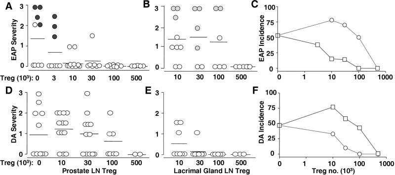
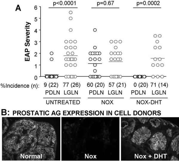
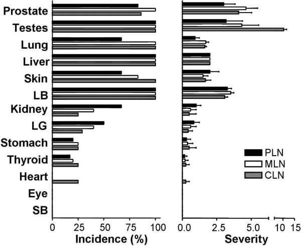
Natural CD4(+)CD25(+)Foxp3(+) regulatory T cells (Treg) effectively prevent autoimmune disease development, but their role in maintaining physiological tolerance against self-Ag of internal organs is not yet defined. In this study, we quantified disease-specific Treg (DS-Treg) as Treg that preferentially suppress one autoimmune disease over another in day 3 thymectomized recipients. A striking difference was found among individual lymph nodes (LN) of normal mice; Treg from draining LN were 15-50 times more efficient than those of nondraining LN at suppressing autoimmune diseases of ovary, prostate, and lacrimal glands. The difference disappeared upon auto-Ag ablation and returned upon auto-Ag re-expression. In contrast, the CD4(+)CD25(-) effector T cells from different individual LN induced multiorgan inflammation with comparable organ distribution. We propose that peripheral tolerance for internal organs relies on the control of autoreactive effector T cells by strategic enrichment of Ag-specific Treg in the regional LN.


Figures

Figure 1
Figure 1
Treg from normal ovarian LN suppress AOD more efficiently than Treg from non-ovarian LN. (A and B): Severity of AOD in d3tx recipients of ovarian LN Treg (A) or non-ovarian LN Treg (B). Filled circles represent positive serum oocyte antibody. (C): AOD incidences between recipients of ovarian LN Treg (squares) and recipients of non-ovarian LN Treg (circles) (n = 6-8).
Figure 2
Figure 2
Selective enrichment of EAP-specific Treg and DA-specific Treg in prostate LN and lacrimal gland LN of normal mice. (A and B): EAP severity between d3tx recipients of Treg from prostate LN (A) and lacrimal gland LN (B). Incidences of EAP between these Treg recipients are shown in C (squares, prostate LN Treg recipients; circles, lacrimal gland LN Treg recipients). (D and E): DA severity between d3tx recipients of Treg from prostate LN (D) and lacrimal gland LN (E). (F): Incidence of DA between these Treg recipients (squares, prostate LN Treg recipients; circles: lacrimal gland LN Treg recipients). Filled circles denote positive prostate autoantibody.
Figure 3
Figure 3
Loss and gain in prostate Ag expression influences EAP-specific Treg activity in the normal prostate LN. (A): EAP severity and incidence in mice that received Treg (0.03 – 0.05×10⁶) from prostate LN (PDLN) or lacrimal gland LN (LGLN), obtained from: normal donors, NOX donors, or donors with NOX and DHT treatment (NOX+DHT). (B): Immunohistochemical detection of prostate antigens in normal mice, NOX mice, and NOX+DHT. P-values determined by Mann-Whitney test.
Figure 4
Figure 4
Organ distribution of tissue inflammation in B6 RAG-/- recipients of CD4+CD25- T cells from individual LN of wild type B6 donors. Data represent the incidence (%) and severity (mean ± SD) of tissue inflammation and destruction. Comparisons are made between pathology induced by lacrimal gland-draining cervical LN (CLN; gray bars), mesenteric LN (MLN; white bars) or prostate LN (PLN; black bars). Each recipient received 0.2×10⁶ CD4+CD25- T cells i.v., and was studied 3-4 weeks later. Data represents the summation of three independent experiments, with two mice per experiment. LB, large bowel; LG, lacrimal gland; SB, small bowel.

References

    1. Scheinecker C, McHugh R, Shevach EM, Germain RN. Constitutive presentation of a natural tissue autoantigen exclusively by dendritic cells in the draining lymph node. J. Exp. Med. 2002;196:1079–1090. - PMC - PubMed
    1. Walker LS, Chodos A, Eggena M, Dooms H, Abbas AK. Antigen-dependent proliferation of CD4+ CD25+ regulatory T cells in vivo. J Exp. Med. 2003;198:249–258. - PMC - PubMed
    1. Fisson S, Darrasse-Jeze G, Litvinova E, Septier F, Klatzmann D, Liblau R, Salomon BL. Continuous activation of autoreactive CD4+ CD25+ regulatory T cells in the steady state. J. Exp. Med. 2003;198:737–746. - PMC - PubMed
    1. Samy ET, Parker LA, Sharp CP, Tung KSK. Continuous control of autoimmune disease by antigen-dependent polyclonal CD4+CD25+ regulatory T cells in the regional lymph node. J. Exp. Med. 2005;202:771–781. - PMC - PubMed
    1. Lathrop SK, Santacruz NA, Pham D, Luo J, Hsieh CS. Antigen-specific peripheral shaping of the natural regulatory T cell population. J. Exp. Med. 2008;205:3105–3117. - PMC - PubMed

Publication types

MeSH terms

Substances