

doi: 10.1107/S1744309109037191.
Epub 2009 Oct 13.
Structure of an N-terminally truncated Nudix hydrolase DR2204 from Deinococcus radiodurans
Affiliations
- PMID: 19923723
- PMCID: PMC2777031
- DOI: 10.1107/S1744309109037191
Item in Clipboard
Structure of an N-terminally truncated Nudix hydrolase DR2204 from Deinococcus radiodurans
Acta Crystallogr Sect F Struct Biol Cryst Commun.
.
Abstract
Nudix pyrophosphatases are a well represented protein family in the Deinococcus radiodurans genome. These hydrolases, which are known to be enzymatically active towards nucleoside diphosphate derivatives, play a role in cleansing the cell pool of potentially deleterious damage products. Here, the structure of DR2204, the only ADP-ribose pyrophosphatase in the D. radiodurans genome that is known to be active towards flavin adenosine dinucleotide (FAD), is presented at 2.0 angstrom resolution.
Figures
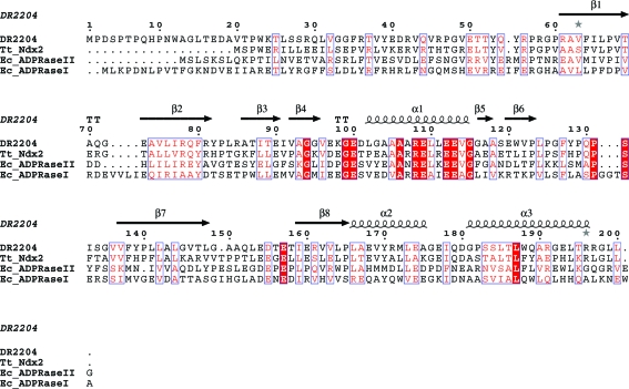
Sequence alignment of DR2204 with the Tt_Ndx2 (Thermus thermophilus HB8; 40% identity), Ec_ADPRaseII (E. coli ORF186; 24% identity) and Ec_ADPRaseI (E. coli ORF209; 21% sequence identity) proteins. Identical residues are shown in white on a red background and similar residues are shown in red on a white background. For the secondary-structure assignment, α-helices are represented as helices, β-strands are represented as arrows and β-turns are marked ‘TT’. This figure was prepared with ESPript (Gouet et al., 1999 ▶). The multiple sequence alignment was performed with CLUSTALW (v1.83) (Thompson et al., 1994 ▶)
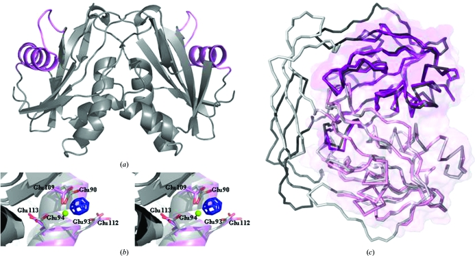
(a) The overall structure of the DR2204 dimer as observed in the asymmetric unit. The protein in grey is shown in cartoon representation, with the Nudix box highlighted in magenta. (b) Stereo picture showing a close-up of the Nudix box. The experimental map (blue; contoured at 10.0σ) calculated from data collected from the gadolinium-derivative crystal used for phasing is overlaid on the final model refined from data collected from the native crystal (magenta). A significant positive peak for the Gd3+ ion is observed in the region where Mg2+ is expected to bind during catalysis, between the side chains of Glu109 and Glu112. Both residues are located in the Nudix-box region, a versatile divalent metal-binding site characteristic of this family of enzymes. For clarity, the model of the ADP-ribose pyrophosphatase (Ndx2) from T. thermophilus (grey; PDB code 2yvm ) including the Mg2+ 1002 ion (in green) bound to residues Glu90 and Glu94 is shown. (c) Superposition of DR2204 (magenta) on the structures of the ADP-ribose pyrophosphatase from T. thermophilus (grey; 0.73 Å r.m.s.d.; PDB code 2yvm ). The surface of DR2204 is overlaid on the proteins represented in ribbon; the dimer formation buries ∼1100 Å2 of solvent-accessible surface. This figure was prepared with PyMOL (DeLano, 2003 ▶).
References
-
- Abrahams, J. P. & Leslie, A. G. W. (1996). Acta Cryst. D52, 30–42. - PubMed
-
- Battista, J. R. (1997). Annu. Rev. Microbiol.51, 203–224. - PubMed
-
- Bessman, M. J., Frick, D. N. & O’Handley, S. F. (1996). J. Biol. Chem.271, 25059–25062. - PubMed
-
- Bhatnagar, S. K. & Bessman, M. J. (1988). J. Biol. Chem.263, 8953–8957. - PubMed
Publication types
MeSH terms
Substances
Associated data
- Actions
LinkOut - more resources
Full Text Sources
