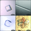
Purification, crystallization and preliminary X-ray diffraction experiments on the breakage-reunion domain of the DNA gyrase from Mycobacterium tuberculosis
- PMID: 19923746
- PMCID: PMC2777054
- DOI: 10.1107/S1744309109042067
Purification, crystallization and preliminary X-ray diffraction experiments on the breakage-reunion domain of the DNA gyrase from Mycobacterium tuberculosis
Abstract
Mycobacterium tuberculosis DNA gyrase, a nanomachine that is involved in the regulation of DNA topology, is the only type II topoisomerase present in this organism and hence is the sole target for fluoroquinolone action. The breakage-reunion domain of the A subunit plays an essential role in DNA binding during the catalytic cycle. Two constructs of 53 and 57 kDa (termed GA53BK and GA57BK) corresponding to this domain have been overproduced, purified and crystallized. Diffraction data were collected from four crystal forms. The resolution limits ranged from 4.6 to 2.7 angstrom depending on the crystal form. The best diffracting crystals belonged to space group C2, with a biological dimer in the asymmetric unit. This is the first report of the crystallization and preliminary X-ray diffraction analysis of the breakage-reunion domain of DNA gyrase from a species containing one unique type II topoisomerase.
Figures


References
-
- Aubry, A., Fisher, L. M., Jarlier, V. & Cambau, E. (2006). Biochem. Biophys. Res. Commun.348, 158–165. - PubMed
-
- Berger, J. M., Gamblin, S. J., Harrison, S. C. & Wang, J. C. (1996). Nature (London), 379, 225–232. - PubMed
-
- Blanc, E., Roversi, P., Vonrhein, C., Flensburg, C., Lea, S. M. & Bricogne, G. (2004). Acta Cryst. D60, 2210–2221. - PubMed
-
- Champoux, J. J. (2001). Annu. Rev. Biochem.70, 369–413. - PubMed
MeSH terms
Substances
Associated data
- Actions
LinkOut - more resources
Full Text Sources
Miscellaneous

