. 2009;14(6):411-22.
doi: 10.1159/000241898. Epub 2009 Nov 16.

Development of a microfluidics-based intracochlear drug delivery device

Affiliations

Development of a microfluidics-based intracochlear drug delivery device

William F Sewell et al. Audiol Neurootol. 2009.

Abstract

Background: Direct delivery of drugs and other agents into the inner ear will be important for many emerging therapies, including the treatment of degenerative disorders and guiding regeneration.

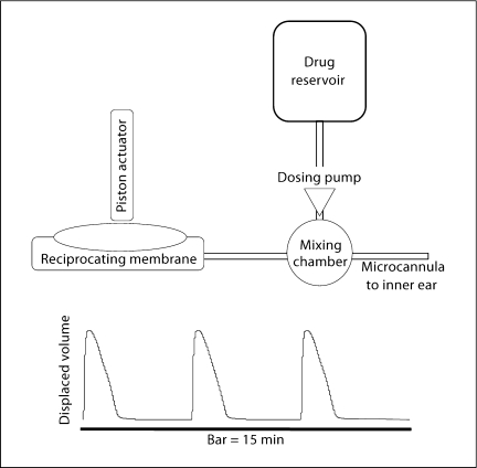
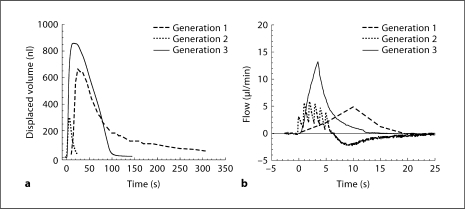
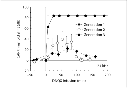
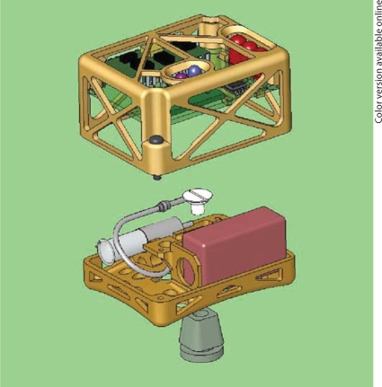
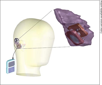
Methods: We have taken a microfluidics/MEMS (MicroElectroMechanical Systems) technology approach to develop a fully implantable reciprocating inner-ear drug-delivery system capable of timed and sequenced delivery of agents directly into perilymph of the cochlea. Iterations of the device were tested in guinea pigs to determine the flow characteristics required for safe and effective delivery. For these tests, we used the glutamate receptor blocker DNQX, which alters auditory nerve responses but not cochlear distortion product otoacoustic emissions.

Results: We have demonstrated safe and effective delivery of agents into the scala tympani. Equilibration of the drug in the basal turn occurs rapidly (within tens of minutes) and is dependent on reciprocating flow parameters.

Conclusion: We have described a prototype system for the direct delivery of drugs to the inner ear that has the potential to be a fully implantable means for safe and effective treatment of hearing loss and other diseases.


Figures

Fig. 1.
Fig. 1.
Schematic representation of the Generation 1 device. We have previously published a thorough description of this device [13]. The device created a reciprocating delivery flow by recirculating perilymph through the system. A fluidic capacitance produced by a long length of compliant tubing produced a delay, measured at the manifold containing the ear cannula, between the positive pressure at the output of the pump and the negative pressure at the input. Placement of a high resistance between the manifold to the microcannula and the capacitor directed fluid out of the microcannula during the initial pulse. Thus, fluid was expelled through the cannula for a short period of time before being withdrawn back into the system.
Fig. 2.
Fig. 2.
Schematic representation of the Generation 2 device. The development of a tunable fluidics capacitor to replace the long length of tubing allowed better specification of the flow characteristics out of the microcannula. For this device, we also developed a microfluidics-based injection port and miniaturized the electronic control to allow the animal to wear the device for periods of days to weeks.
Fig. 3.
Fig. 3.
Schematic representation of the Generation 3 device. In this device, the reciprocating delivery is achieved by a ‘push-pull’ fluid flow. In the initial benchtop experiments described in this paper, a syringe pump (Harvard Apparatus PHD2000) was substituted for the reciprocating membrane and piston actuator.
Fig. 4.
Fig. 4.
Displaced fluid volume (a) and flow (b) as a function of time for each of the 3 devices. Displaced volume represents the volume of fluid injected into and then withdrawn from the inner ear. A positive flow indicates flow from the device to the cochlea and a negative flow is the return from the cochlea to the device. All 3 devices have both positive and negative flow components, though only the generation 2 device entered the negative flow mode during the time indicated in b (25 s). In both the generation 1 and generation 2 devices, volume is displaced in each cycle by a closely spaced series of pump pulses. The capacitance properties of the generation 1 system integrate those closely spaced pump pulses more smoothly than the generation 2 device. In the generation 3 device, inflow and outflow are mediated by a positive and negative volume displacement via a ‘push-pull’ pump.
Fig. 5.
Fig. 5.
CAP threshold shift, measured 1 h after DNQX infusion, is plotted as a function of tone-pip frequency. Cochlear regions near the base respond best to high-frequency tones. CAP threshold shift is related to the concentration of DNQX in each cochlear region. The 3 plots represent changes in CAP (means ± SE) associated with the 3 devices. The flow and volume parameters for each device are indicated in figure 4. For each device, flow and volume were optimized in a few initial experiments before settling on the parameters indicated in figure 4.
Fig. 6.
Fig. 6.
Different reciprocating profiles of the 3 generations of our device produced differences in the rapidity and efficacy of drug effects. Data are means ± SE.
Fig. 7.
Fig. 7.
Solidworks diagram of the miniaturized wearable device. The Wilson-Greatbatch pump, the microfluidics components, and the battery are placed on the lower level, while the upper level contains the microprocessor and the communications circuitry.
Fig. 8.
Fig. 8.
Concept illustration of the device. The pump, fluid reservoir, and electronics are to be placed in space created in the mastoid. An external charger and control module will allow modification of the dose regimen and the drug delivery sequence.

Similar articles

Cited by

References

    1. Holley MC. Application of new biological approaches to stimulate sensory repair and protection. Br Med Bull. 2002;63:157–169. - PubMed
    1. Ryan AF, Harris JP, Keithley EM. Immune-mediated hearing loss: basic mechanisms and options for therapy. Acta Otolaryngol Suppl. 2002;548:38–43. - PubMed
    1. Shinohara T, Bredberg G, Ulfendahl M, Pyykko I, Olivius NP, Kaksonen R, Lindstrom B, Altschuler R, Miller JM. Neurotrophic factor intervention restores auditory function in deafened animals. Pro Natl Acad Sci USA. 2002;99:1657–1660. - PMC - PubMed
    1. Raphael Y. Cochlear pathology, sensory cell death and regeneration. Br Med Bull. 2002;63:25–38. - PubMed
    1. Lalwani AK, Jero J, Mhatre AN. Developments in cochlear gene therapy. Adv Otorhinolaryngol. 2002;61:28–33. - PubMed

Publication types