. 2009 Oct-Dec;3(4):224-35.
doi: 10.4161/pri.3.4.10112. Epub 2009 Oct 16.

Amyloid fibrils of human prion protein are spun and woven from morphologically disordered aggregates

Affiliations

Amyloid fibrils of human prion protein are spun and woven from morphologically disordered aggregates

Karin Almstedt et al. Prion. 2009 Oct-Dec.

Abstract

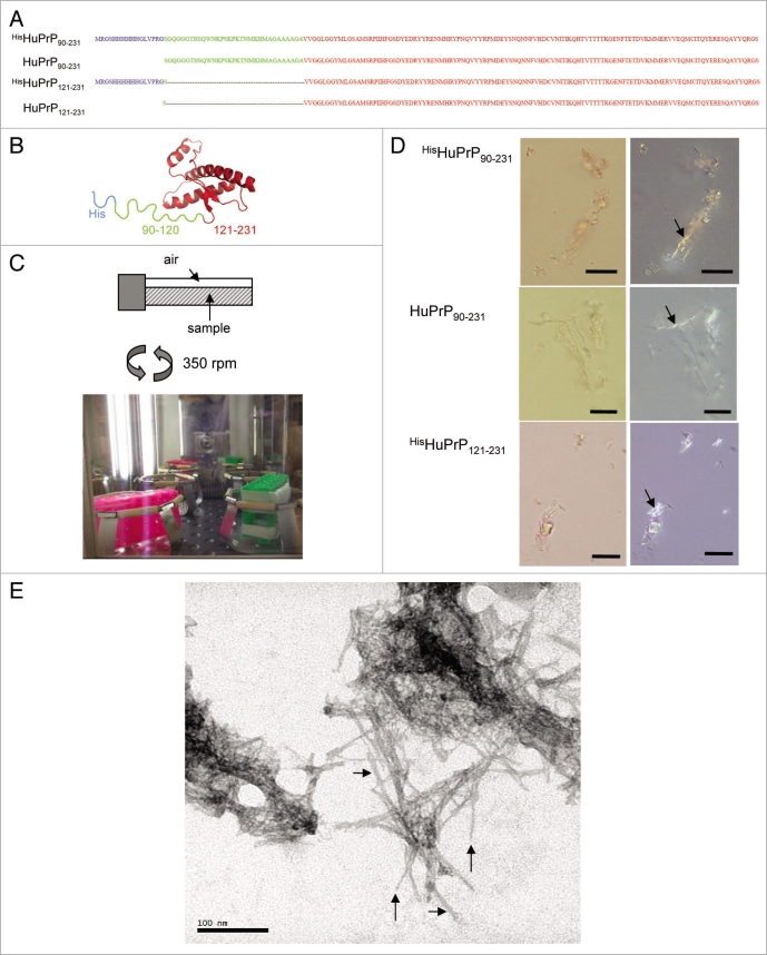
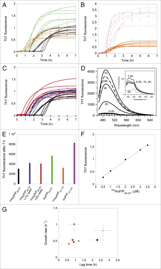
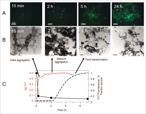
Propagation and infectivity of prions in human prionopathies are likely associated with conversion of the mainly alpha-helical human prion protein, HuPrP, into an aggregated form with amyloid-like properties. Previous reports on efficient conversion of recombinant HuPrP have used mild to harsh denaturing conditions to generate amyloid fibrils in vitro. Herein we report on the in vitro conversion of four forms of truncated HuPrP (sequences 90-231 and 121-231 with and without an N-terminal hexa histidine tag) into amyloid-like fibrils within a few hours by using a protocol (phosphate buffered saline solutions at neutral pH with intense agitation) close to physiological conditions. The conversion process monitored by thioflavin T, ThT, revealed a three stage process with lag, growth and equilibrium phases. Seeding with preformed fibrils shortened the lag phase demonstrating the classic nucleated polymerization mechanism for the reaction. Interestingly, comparing thioflavin T kinetics with solubility and turbidity kinetics it was found that the protein initially formed nonthioflavionophilic, morphologically disordered aggregates that over time matured into amyloid fibrils. By transmission electron microscopy and by fluorescence microscopy of aggregates stained with luminescent conjugated polythiophenes (LCPs); we demonstrated that HuPrP undergoes a conformational conversion where spun and woven fibrils protruded from morphologically disordered aggregates. The initial aggregation functioned as a kinetic trap that decelerated nucleation into a fibrillation competent nucleus, but at the same time without aggregation there was no onset of amyloid fibril formation. The agitation, which was necessary for fibril formation to be induced, transiently exposes the protein to the air-water interface suggests a hitherto largely unexplored denaturing environment for prion conversion.


Figures

Figure 1
Figure 1
Sequences of HuPrP variants and aggregation into amyloid fibrils. (A) Primary sequences of the HuPrP variants: HisHuPrP90–231, HuPrP90–231, HisHuPrP121–231 and HuPrP121–231. The color coding show the 17 residue His-tag sequence in blue, segment 90–120 in green and the folded domain 121–231 in red. (B) Structure of HisHuPrP90–231 with the same color coding as in (A). The structure of the folded domain was generated in PyMol using the pdb entry code 1QLX. (C) Sample set up for fibril formation conditions. Screw-on-cap test tubes (2 mL) were filled with 800 µL sample and were placed in a rack tilted vertically, to afford horizontally aligned samples providing a large air-water interface (top image). The rack was placed in a shaking incubator inducing 350 rpm orbital shaking at 37°C. (D) Micrographs showing open (left) and crossed polarizers (right) of Congo red stained HuPrP aggregates incubated 24 h under fibrillation conditions. Arrows indicate green birefringent aggregates. Scale bars indicate 10 µm. (E) Transmission electron micrograph of HisHuPrP90–231 fibrils (24 h incubation) stained with uranyl acetate taken at 250,000 fold magnification showing needle like amyloid fibrils. Vertical arrows show 3 nm wide filaments and horizontal arrows indicate 6 nm wide fibrils. Scale bar indicate 100 nm.
Figure 2
Figure 2
Fibril formation kinetics of HuPrP variants monitored by ThT fluorescence. (A) Representative fibril formation kinetic traces of HisHuPrP90–231 (black), HuPrP90–231 (green) and HisHuPrP121–231 (orange). Fibril formation was performed at 37°C with continuous shaking (350 rpm) of 12 µM protein in buffer F: 100 mM NaCl, 50 mM KCl, 50 mM phosphate, pH 7.3 Assay conditions (aliquots from the above samples) were performed in buffer F containing 2 µM ThT and 2 µM final concentration of protein. (B) Representative fibril formation kinetic traces of HisHuPrP121–231 (orange), HuPrP121–231 (magenta). Fibrillation and assay conditions as in (A). (C) Representative fibril formation kinetic traces of unseeded HisHuPrP90–231 (black), with 1% seed (blue) and 5% seed (red) of preformed sonicated HisHuPrP90–231 fibrils. Fibrillation and assay conditions as in (A). The open triangles (dashed line) show the trace for HisHuPrP90–231 under identical conditions as in the unseeded samples under quiescent (non-shaken) settings. (D) Representative fluorescence spectra of ThT (2 µM) during fibril assay conditions mixed with aliquots of HisHuPrP90–231 (final concentration of 2 µM) at different time points of fibrillation (indicated). The inset shows the zoomed in fluorescence spectra during the lag phase, indicating a slight increase in fluorescence >0 h. The ThT fluorescence spectrum at 24 h is shown with a dashed line. (E) Bar plot of the average final ThT fluorescence at 7 h of fibrillation. Color code as in (A–C). (F) Titration curve of preformed (7 h) amyloid fibrils of HisHuPrP90–231 (concentration given on a monomer basis) against a fixed concentration of ThT (2 µM), showing a linear dependence of fluorescence intensity versus the concentration of fibrils. (G) Correlation diagram of average lag phases versus average growth rates of the different samples. Unseeded HisHuPrP90–231 at 12 µM (black circle), n = 15; Unseeded HisHuPrP90–231 at 6 µM (black open circle), n = 10; 1% seed HisHuPrP90–231 at 12 µM (blue circle), n = 9; 5% seed HisHuPrP90–231 at 12 µM (red circle), n = 5; Unseeded HuPrP90–231 at 12 µM (green circle), n = 7; Unseeded HisHuPrP121–231 at 12 µM (orange circle), n = 6; and Unseeded HuPrP121–231 at 12 µM (magenta circle), n = 8. Error bars indicate standard deviations and n present the number of identical samples run in the fibrillation assay for each variant and condition.
Figure 3
Figure 3
Morphology of HisHuPrP90–231 aggregates during fibrillation. (A) Fluorescence micrographs of pelleted ThT stained aggregates at different time points, showing intense ThT fluorescence at 5 h and 24 h, but not at earlier timepoints. Images were collected using the 470/40 nm bandpass filter (LP515). Scale bars indicate 50 µm. (B) Transmission electron microscopy images of negatively stained aggregates at different time points taken at 200,000 fold magnification, showing the conversion of aggregates with apparent morphological disorder that mature towards fibrils over time. Scale bar represents 100 nm. (C) Kinetic traces of turbidity at 330 nm (average trace of three samples) (red open squares), protein solubility (black closed squares) and the fitted ThT fluorescence (average trace of 15 samples) (black dashed trace), run under identical conditions.
Figure 4
Figure 4
Amyloid fibrils are spun and woven from disordered aggregates. (A) PK digestion of HisHuPrP90–231 at different time points during the fibril formation reaction. Recombinant HisHuPrP90–231 (0.2 mg/ml) was treated with PK at a PK/PrP ratio of 1:100 at room temperature for 0–60 min and digestion was stopped by boiling aliquots in SDS loading buffer prior to loading the SDS-PAGE gel. Proteins were visualized using coomassie. (B) Fluorescence spectral distributions of self-sedimented HisHuPrP90–231 aggregates (after 15 min and 24 h of fibrillation) stained with the LCP PTAA (at pH 7.5). The ratio plot display fluorescence intensity ratios at 531 nm/590 nm versus 531 nm/641 nm of five individual regions of interest of four images for each time point. Both types of aggregates showed different distributions of emission spectra, where the average spectra of early aggregates were significantly green shifted compared to the 24 h fibrils, indicating a more planar and stacked conformation of the latter aggregated state. Fluorescence micrographs of representative aggregates using both pseudo-color (“filter view”) (where green color denotes 500–600 nm and red color shows fluorescence at 600–700 nm) and computer generated “real view” images are shown to the right. (C) Pseudo-color fluorescence micrographs (right) of self-sedimented HisHuPrP90–231 fibrils (24 h) stained with the LCP tPTT (at pH 4.0), showing the concomitant presence of at least two conformations (green and red fluorescence as in B) of the bound LCP indicated with blue vertical arrow and red horizontal arrow. The left panel shows the fluorescence spectra from these species. Image and spectral analysis was performed using a SpectraCube® (Optical head) module (ASI, Israel), through a 470/40 nm bandpass filter (LP515). (D) Transmission electron micrograph of HisHuPrP90–231 aggregates (formed during agitation for four days in 50 mM phosphate buffer pH 7.3 at 37°C) stained with uranyl acetate taken at 60,000 fold magnification showing an area of interwoven amyloid fibrils flanked by regions of apparent disordered aggregates. Scale bar indicates 200 nm.

Similar articles

Cited by

References

    1. Westermark P. Aspects on human amyloid forms and their fibril polypeptides. The FEBS journal. 2005;272:5942–5949. - PubMed
    1. Chiti F, Dobson CM. Protein misfolding, functional amyloid and human disease. Ann Rev Biochem. 2006;75:333–366. - PubMed
    1. Mishra R, Sorgjerd K, Nystrom S, Nordigarden A, Yu YC, Hammarstrom P. Lysozyme amyloidogenesis is accelerated by specific nicking and fragmentation but decelerated by intact protein binding and conversion. J Mol Biol. 2007;366:1029–1044. - PubMed
    1. Liberski PP. Amyloid plaques in transmissible spongiform encephalopathies (prion diseases) Folia neuropathologica. 2004;42:109–119. - PubMed
    1. Prusiner SB. Novel proteinaceous infectious particles cause scrapie. Science. 1982;216:136–144. - PubMed

Publication types

LinkOut - more resources