

Novel xenograft and cell line derived from an invasive intraductal papillary mucinous neoplasm of the pancreas give new insights into molecular mechanisms
- PMID: 19924021
- PMCID: PMC3086384
- DOI: 10.1097/MPA.0b013e3181bd5c10
Novel xenograft and cell line derived from an invasive intraductal papillary mucinous neoplasm of the pancreas give new insights into molecular mechanisms
Abstract
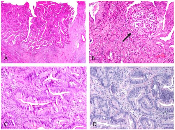
Objectives: Intraductal papillary mucinous neoplasms (IPMNs) of the pancreas are a unique entity with malignant potential. Histologically, pancreatic ductal adenocarcinoma (PDAC) arising in IPMN (intraductal papillary mucinous carcinoma [IPMC]) appears similar to sporadic PDAC; biologically, however, IPMC seems to have a less aggressive clinical course. Little is known about the genetic signature of IPMC. In this study, we describe a novel xenograft model and cell culture created to biologically and genetically characterize these tumors.
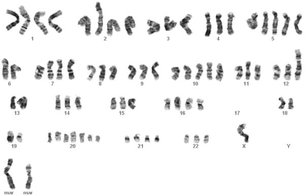
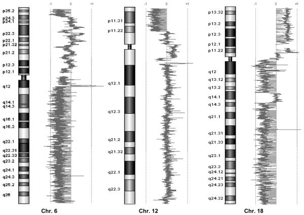
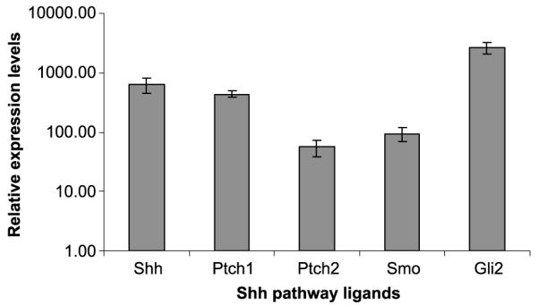
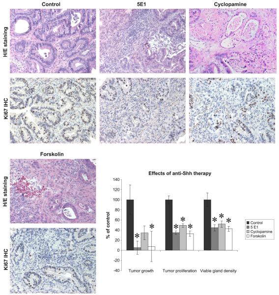
Methods: Xenograft mice and cell lines were created from IPMC. Global genomic changes were evaluated by cytogenetic analysis and array comparative genomic hybridization. Specific mutations and sonic hedgehog (Shh) pathway activity were examined and xenografts evaluated for sensitivity to anti-Shh therapy.
Results: Cytogenetic analysis showed a tetraploid karyotype with multiple aberrations. KRAS and p53 mutations and overexpression of the Shh pathway were identified. Array comparative genomic hybridization revealed multiple chromosomal aberrations comparable with previously published data in IPMNs. Murine xenograft tumors were sensitive to anti-Shh treatment.
Conclusions: Characterization of IPMC cell lines and xenografts reveals similarities to previously published data on IPMN. In comparison to PDAC, moreover, these data reveal shared aberrations and distinct genomic changes. Thus, these xenograft model and cell lines may be useful for future preclinical investigations.
Figures
Similar articles
-
Disease spectrum of intraductal papillary mucinous neoplasm with an associated invasive carcinoma invasive IPMN versus pancreatic ductal adenocarcinoma-associated IPMN.Pancreas. 2013 Nov;42(8):1267-74. doi: 10.1097/mpa.0b013e3182954137. Pancreas. 2013. PMID: 24308063
-
BRAF and KRAS gene mutations in intraductal papillary mucinous neoplasm/carcinoma (IPMN/IPMC) of the pancreas.Cancer Lett. 2007 May 8;249(2):242-8. doi: 10.1016/j.canlet.2006.09.007. Epub 2006 Nov 9. Cancer Lett. 2007. PMID: 17097223 Free PMC article.
-
Intraductal papillary mucinous neoplasms of the pancreas with distinct pancreatic ductal adenocarcinomas are frequently of gastric subtype.Ann Surg. 2013 Jul;258(1):141-51. doi: 10.1097/SLA.0b013e31828cd008. Ann Surg. 2013. PMID: 23532108
-
Intraductal Papillary Mucinous Carcinoma Versus Conventional Pancreatic Ductal Adenocarcinoma: A Comprehensive Review of Clinical-Pathological Features, Outcomes, and Molecular Insights.Int J Mol Sci. 2021 Jun 23;22(13):6756. doi: 10.3390/ijms22136756. Int J Mol Sci. 2021. PMID: 34201897 Free PMC article. Review.
-
Intraductal papillary mucinous carcinoma versus pancreatic ductal adenocarcinoma: A systematic review and meta-analysis.Int J Surg. 2019 Nov;71:91-99. doi: 10.1016/j.ijsu.2019.09.014. Epub 2019 Sep 20. Int J Surg. 2019. PMID: 31546033
Cited by
-
Intraductal papillary mucinous tumors of the pancreas: biology, diagnosis, and treatment.Oncologist. 2010;15(12):1294-309. doi: 10.1634/theoncologist.2010-0151. Epub 2010 Dec 8. Oncologist. 2010. PMID: 21147870 Free PMC article. Review.
-
Intraductal papillary mucinous neoplasm of the pancreas rapidly xenografts in chicken eggs and predicts aggressiveness.Int J Cancer. 2018 Apr 1;142(7):1440-1452. doi: 10.1002/ijc.31160. Epub 2017 Nov 29. Int J Cancer. 2018. PMID: 29143337 Free PMC article.
-
Epithelial to Stromal Re-Distribution of Primary Cilia during Pancreatic Carcinogenesis.PLoS One. 2016 Oct 26;11(10):e0164231. doi: 10.1371/journal.pone.0164231. eCollection 2016. PLoS One. 2016. PMID: 27783689 Free PMC article.
-
Diversity of Precursor Lesions For Pancreatic Cancer: The Genetics and Biology of Intraductal Papillary Mucinous Neoplasm.Clin Transl Gastroenterol. 2017 Apr 6;8(4):e86. doi: 10.1038/ctg.2017.3. Clin Transl Gastroenterol. 2017. PMID: 28383565 Free PMC article.
-
Clinical and Pre-Clinical Evidence of Carbonic Anhydrase IX in Pancreatic Cancer and Its High Expression in Pre-Cancerous Lesions.Cancers (Basel). 2020 Jul 22;12(8):2005. doi: 10.3390/cancers12082005. Cancers (Basel). 2020. PMID: 32707920 Free PMC article. Review.
References
-
- Conlon KC. Intraductal papillary mucinous tumors of the pancreas. J Clin Oncol. 2005;23:4518–4523. - PubMed
-
- Sarr MG, Murr M, Smyrk TC, et al. Primary cystic neoplasms of the pancreas. Neoplastic disorders of emerging importance-current state-of-the-art and unanswered questions. J Gastrointest Surg. 2003;7:417–428. - PubMed
Publication types
MeSH terms
Substances
Grants and funding
LinkOut - more resources
Full Text Sources
Medical
Research Materials
Miscellaneous
