. 2009 Oct 26;14(11):4300-11.
doi: 10.3390/molecules14114300.

Silibinin suppresses TNF-alpha-induced MMP-9 expression in gastric cancer cells through inhibition of the MAPK pathway

Affiliations

Silibinin suppresses TNF-alpha-induced MMP-9 expression in gastric cancer cells through inhibition of the MAPK pathway

Sangmin Kim et al. Molecules. .

Abstract

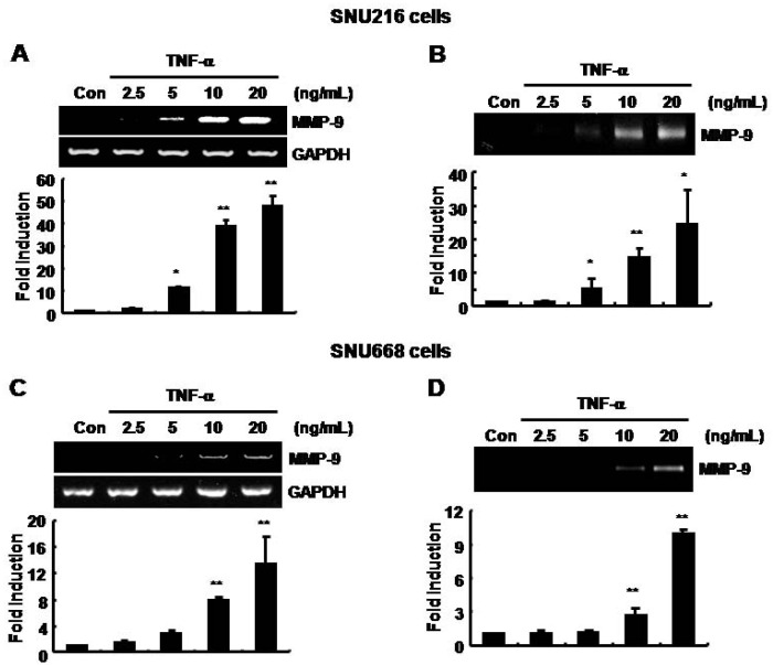
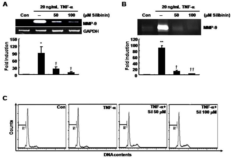
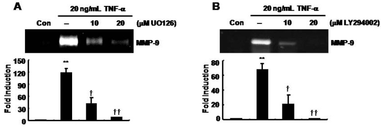
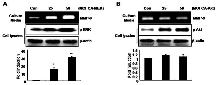
Tumor necrosis factor (TNF)-alpha is one of the pro-inflammatory cytokines highly expressed in Helicobacter pylori that inhibits gastric acid secretion. In this study we determined the effect of silibinin on TNF-alpha-induced MMP-9 expression in gastric cancer cell lines. MMP-9 mRNA and protein expression was dose-dependently increased by TNF-alpha in SNU216 and SNU668 gastric cancer cells. On the other hand, TNF-alpha-induced MMP-9 expression was dose-dependently suppressed by silibinin. To verify the regulatory mechanism of silibinin on TNF-alpha-induced MMP-9 expression, the gastric cancer cell lines were pretreated with silibinin prior to TNF-alpha. TNF-alpha-induced MMP-9 expression was inhibited by the MEK1/2 specific inhibitor, UO126. Finally, we investigated the effect of adenoviral constitutively active (CA)-MEK and CA-Akt on MMP-9 expression. The expression of MMP-9 was significantly increased by CA-MEK overexpression, but not by CA-Akt overexpression. Taken together, we suggest that silibinin down-regulates TNF-alpha- induced MMP-9 expression through inhibition of the MEK/ERK pathway in gastric cancer cells.


Figures

Figure 1
Figure 1
The basal levels of MMP-9 mRNA and protein expression were increased by TNF-α in a dose-dependent manner in SNU216 and SNU668 gastric cancer cells. After serum-starvation for 24 h, cells were treated with TNF-αat the concentrations indicated for 24 h in fresh serum-free media. (A, C) The level of MMP-9 mRNA expression was analyzed by RT-PCR in SNU216 and SNU668 gastric cancer cells. (B, D) The level of MMP-9 protein expression was analyzed by zymography in SNU216 and SNU668 gastric cancer cells. These results are representative of three independent experiments. The values shown are the mean ± SEM. * P < 0.05, ** P < 0.01 vs. control. Con; control.
Figure 2
Figure 2
TNF-α-induced MMP-9 mRNA and protein expression was decreased by silibinin in a dose-dependent manner in SNU216 gastric cancer cells. After serum-starvation for 24 h, cells were pretreated with silibinin at the concentrations indicated for 60 min, and then treated with 20 μg/mL of TNF-α for 24 h. MMP-9 mRNA and protein expression were analyzed by RT-PCR (A) and zymography (B), respectively. (C) Under the same conditions, the cell cycle was analyzed by FACS analysis, as described in the Materials and Methods. These results are representative of three independent experiments. The values shown are the mean ± SEM. * P < 0.05, ** P < 0.01 vs. control, † P < 0.05, †† P < 0.01 vs. TNF-α-treated cells. Con; control.
Figure 3
Figure 3
TNF-α-induced MMP-9 expression was decreased by UO126 and LY294002 in SNU216 gastric cancer cells. After serum-starvation for 24 h, cells were pretreated with UO126 (A) and LY294002 (B) at the indicated concentrations for 30 min and then treated with 20 μg/mL TNF-α for 24 h. MMP-9 gelatinase activity was analyzed in culture media by zymography. These results are representative of three independent experiments. The values shown are the mean ± SEM. ** P < 0.01 vs. control, † P < 0.05, †† P < 0.01 vs. TNF-α-treated cells. Con; control.
Figure 4
Figure 4
TPA-induced MMP-9 expression is increased by Ad-CA-MEK, but not by Ad-CA-Akt, in SNU216 gastric cancer cells. After Ad-CA-MEK (A) and CA-Akt (B) transfection for 24 h, cells were incubated in serum-free media for 24 h and then fresh serum-free media was added for 24 h. MMP-9 protein expression was analyzed in culture media by zymography. Using the cell lysates, the phosphorylation of ERK (A) and Akt (B) was analyzed by Western blotting. These results are representative of three independent experiments. The values shown are the mean ± SEM. * P < 0.05, ** P < 0.01 vs. control. Con; control.

References

    1. Singh R.P., Agarwal R. Flavonoid antioxidant silymarin and skin cancer. Antioxid Redox Signal. 2002;4:655–663. doi: 10.1089/15230860260220166. - DOI - PubMed
    1. Kim S., Choi J.H., Lim H.I., Lee S.K., Kim W.W., Kim J.S., Kim J.H., Choe J.H., Yang J.H., Nam S.J., Lee J.E. Silibinin prevents TPA-induced MMP-9 expression and VEGF secretion by inactivation of the Raf/MEK/ERK pathway in MCF-7 human breast cancer cells. Phytomedicine. 2009;16:573–580. doi: 10.1016/j.phymed.2008.11.006. - DOI - PubMed
    1. Sharma G., Singh R.P., Chan D.C., Agarwal R. Silibinin induces growth inhibition and apoptotic cell death in human lung carcinoma cells. Anticancer Res. 2003;23:2649–2655. - PubMed
    1. Agarwal R., Agarwal C., Ichikawa H., Singh R.P., Aggarwal B.B. Anticancer potential of silymarin: from bench to bed side. Anticancer Res. 2006;26:4457–4498. - PubMed
    1. Locksley R.M., Killeen N., Lenardo M.J. The TNF and TNF receptor superfamilies: Integrating mammalian biology. Cell. 2001;104:487–501. doi: 10.1016/S0092-8674(01)00237-9. - DOI - PubMed

Publication types

MeSH terms