. 2009 Dec 17;462(7275):880-5.
doi: 10.1038/nature08616. Epub 2009 Nov 18.

Transport mechanism of a bacterial homologue of glutamate transporters

Affiliations

Transport mechanism of a bacterial homologue of glutamate transporters

Nicolas Reyes et al. Nature. .

Abstract

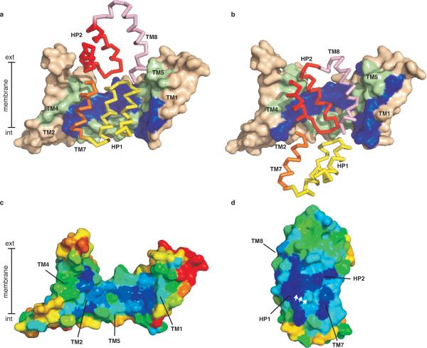
Glutamate transporters are integral membrane proteins that catalyse a thermodynamically uphill uptake of the neurotransmitter glutamate from the synaptic cleft into the cytoplasm of glia and neuronal cells by harnessing the energy of pre-existing electrochemical gradients of ions. Crucial to the reaction is the conformational transition of the transporters between outward and inward facing states, in which the substrate binding sites are accessible from the extracellular space and the cytoplasm, respectively. Here we describe the crystal structure of a double cysteine mutant of a glutamate transporter homologue from Pyrococcus horikoshii, Glt(Ph), which is trapped in the inward facing state by cysteine crosslinking. Together with the previously determined crystal structures of Glt(Ph) in the outward facing state, the structure of the crosslinked mutant allows us to propose a molecular mechanism by which Glt(Ph) and, by analogy, mammalian glutamate transporters mediate sodium-coupled substrate uptake.


Figures

Figure 1
Figure 1. Cross-linking of GltPh-K55C/A364C
a, Cartoon representation of two substrate-bound GltPh protomers (PDB code 2NWX) viewed in the membrane plain. The third protomer and TM4 of the protomer on the right are removed for clarity. TMs 1 through 6 are coloured salmon and wheat; the carboxyl terminal cores are light blue; and the hairpins HP1 and HP2 are dark blue. Bound L-asp and Na+ are shown as sticks and purple spheres, respectively. Orange spheres correspond to the Cα atoms of residues 55 and 364. All molecular representations have been generated using Pymol. b, SDS PAGE analysis of GltPh-K55C/A364C before and after incubation with 100 μM CuPhen for indicated periods of time. Detergent-solubilized purified GltPh-K55C/A364C (left) and unpurified transporter in crude E. coli membranes (right) were visualized by Coomassie staining and western blotting, respectively. c, Cross-linking of GltPh-K55C/A364C in the presence of 50 μM HgCl2. Samples were analyzed as in b.
Figure 2
Figure 2. GltPh-55C/364CHg in the inward facing substrate-bound state
Cartoon representation of the single protomers (a) and surface representation of the trimers sliced through the binding sites (b). WT GltPh and GltPh-55C/364CHg are shown on the left and right, respectively. In (a), TMs 1, 2, 4 and 5 are coloured wheat with the remainder of the protomer light blue. Na+ are shown as purple spheres. c, Extracellular view of GltPh-55C/364CHg with straight and curved lines delineating individual protomers and transport domains, respectively. The trimerization domains (wheat) and transport domains (blue) are connected by short cytoplasmic (solid arrows) and extracellular (open arrows) loops, highlighted in red. The long TM3–4 loops (simple arrows) cross over the transport domains. d, Isothermal titration calorimetry analysis of L-asp binding to GltPh-55C/364CHg. Shown are the binding heats in 10 mM NaCl. The linear dependence (slope = −2.6 ± 0.7) of the log of the apparent dissociation constant, Kd, on the log of Na+ concentration is shown in the inset. GltPh-55C/364CHg was exchanged into Na+/L-asp-free buffer, diluted to 15–20 μM in the reaction cell of the Microcal ITC200, supplemented with indicated concentrations of Na+, and titrated with L-asp at 25 °C. The binding enthalpy and the apparent number of binding sites were 23.6 ± 0.8 kcal/mol and 0.4 ± 0.03 (n=6), respectively.
Figure 3
Figure 3. Domain interaction surfaces
Trimerization domains of WT GltPh (a) and GltPh-55C/364CHg (b) are shown in surface representation and coloured wheat. Residues involved in domain contacts, identified by ProFace server, are coloured blue (TM1 and 2) and green (TM4 and 5). Interacting structural elements of the transport domain are shown in ribbon representation: HP1 (yellow) and TM7 (orange) in WT GltPh, and HP2 (red) and TM8 (pink) in GltPh-55C/364CHg. Surface representation of the trimerization (c) and transport (d) domains coloured according to evolutionary conservation. Dark blue and red correspond to the highly conserved and variable residues, respectively. The interacting surfaces are facing the viewer and the white arrows mark the highly conserved serine-rich signature motif in HP1. Conservation scores were calculated using Consurf server and 212 SLC1 sequences with less than 60 % identity harvested from Pfam database and aligned in ClustalW2.
Figure 4
Figure 4. Amino-terminal inverted structural repeat
Cartoon representation of TMs 1–3 (blue) and TMs 4–6 (green) in the GltPh-55C/364CHg viewed in the membrane plain (a), and their structural superposition (b). c, Symmetrical helices, TMs 1–2 and TMs 4–5, form the interaction surface within the transport domain, which is partitioned into intracellular and extracellular halves delineated by the dotted line. (d) Schematic representation of GltPh trimerization (orange) and the transport (light blue) domains. Two inverted structural repeats are emphasized by blue and green, and yellow and red trapezoids. Structure of TM2–3 and TM5–6 loops in WT GltPh (e) and GltPh-55C/364CHg (f). TMs 2 through 6 are shown in cartoon representation with 4 omitted for clarity. The transporter core is shown in transparent surface representation. Bound L-asp and the highly conserved glycines are shown as spheres.
Figure 5
Figure 5. Schematic transport mechanism
Shown is a single transporter protomer. Substrate and sodium binding to the outward and inward facing states is coupled to the closure of the extracellular and intracellular gates, HP2 (red) and HP1 (yellow), respectively. Isomerization between the outward and inward facing occluded states occurs upon movement of the transport domain (blue), relative to the trimerization domain (grey). The inward facing open state has not been structurally characterized and is hypothetical.

References

    1. Danbolt NC. Glutamate uptake. Prog Neurobiol. 2001;65:1–105. - PubMed
    1. Saier MH, Jr., Tran CV, Barabote RD. TCDB: the Transporter Classification Database for membrane transport protein analyses and information. Nucleic Acids Res. 2006;34:D181–6. - PMC - PubMed
    1. Mitchell P. A general theory of membrane transport from studies of bacteria. Nature. 1957;180:134–6. - PubMed
    1. Patlak CS. Contributions to the theory of active transport: II. The gate type non-carrier mechanism and generalizations concerning tracer flow, efficiency, and measurement of energy expenditure. Bulletin of Mathematical Biology. 1957;19:209–235.
    1. Jardetzky O. Simple allosteric model for membrane pumps. Nature. 1966;211:969–70. - PubMed

Publication types

MeSH terms

Associated data