. 2009 Nov 19;462(7271):358-62.
doi: 10.1038/nature08575.

Systems-level dynamic analyses of fate change in murine embryonic stem cells

Affiliations

Systems-level dynamic analyses of fate change in murine embryonic stem cells

Rong Lu et al. Nature. .

Abstract

Molecular regulation of embryonic stem cell (ESC) fate involves a coordinated interaction between epigenetic, transcriptional and translational mechanisms. It is unclear how these different molecular regulatory mechanisms interact to regulate changes in stem cell fate. Here we present a dynamic systems-level study of cell fate change in murine ESCs following a well-defined perturbation. Global changes in histone acetylation, chromatin-bound RNA polymerase II, messenger RNA (mRNA), and nuclear protein levels were measured over 5 days after downregulation of Nanog, a key pluripotency regulator. Our data demonstrate how a single genetic perturbation leads to progressive widespread changes in several molecular regulatory layers, and provide a dynamic view of information flow in the epigenome, transcriptome and proteome. We observe that a large proportion of changes in nuclear protein levels are not accompanied by concordant changes in the expression of corresponding mRNAs, indicating important roles for translational and post-translational regulation of ESC fate. Gene-ontology analysis across different molecular layers indicates that although chromatin reconfiguration is important for altering cell fate, it is preceded by transcription-factor-mediated regulatory events. The temporal order of gene expression alterations shows the order of the regulatory network reconfiguration and offers further insight into the gene regulatory network. Our studies extend the conventional systems biology approach to include many molecular species, regulatory layers and temporal series, and underscore the complexity of the multilayer regulatory mechanisms responsible for changes in protein expression that determine stem cell fate.


Figures

Figure 1
Figure 1. Measuring changes in the epigenome, the transcriptome and the nuclear proteome after Nanog downregulation
a, Experimental design. AP, alkaline phosphatase; IP, immunoprecipitation; iTRAQ, isobaric tag for relative and absolute quantification; MS, mass spectrometry. b, The lentiviral vector construct to conditionally regulate Nanog expression levels. dLTR, deleted long-terminal repeat; FLAP, nucleotide segment that improves transduction efficiency; Teton, tetracycline transactivator; WRE, woodchuck hepatitis virus post-transcriptional regulatory element. c, Efficacy of Nanog protein downregulation as measured by mass spectrometry (bar chart) and western blot (image, bottom). Error bars denote the s.d. of duplicate measurements. d, Summary of the numbers of genes with significant changes at different molecular layers on each day. Increased and decreased levels are shown in orange and green, respectively.
Figure 2
Figure 2. Comparisons across different molecular regulatory layers
a, Proteins with significant changes on each day are assigned to one of four categories on the basis of concordance between expression steps (Methods). The percentages on the left are calculated according to the number of proteins in each category. The P-value bar on the right gives the inclusion significance level. b, Examples of proteins from each of the four categories. Black dots represent the exact values for each experimental replicate. c, Selected gene-ontology (GO) categories that are overrepresented at each gene expression step. The complete panel is shown in Supplementary Fig. 5.
Figure 3
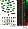
Figure 3. Dynamic changes in ESC networks
a, The core ESC protein–protein interaction network (connections) overlaid with dynamic protein changes observed in our data (rectangles are divided into three segments representing changes on days 1, 3 and 5 compared to day 0). b, Heat map of multimolecular layer gene expression changes for Nanog-binding genes. Shown are the genes whose data were obtained with high confidence on all four molecular layers. Genes are ranked on the basis of changes in protein levels. c, The pluripotency transcriptional regulatory network (arrows) overlaid with mRNA fold changes (colours) from our data.
Figure 4
Figure 4. Interactive visualization of the multilayer dynamic data
a, Snapshots from heat map movies showing 400 genes with the most significant changes in protein levels on day 5. The position (pixel) of each gene locus is the same in all 12 heat maps. b, Snapshots from dynamic scatter plots illustrating concurrent changes in mRNAs and proteins. Red dots represent genes that have been identified to have important roles in ESCs. Supplementary Fig. 8 and the website http://amp.pharm.mssm.edu/ronglu are interactive and each gene can be displayed as a line plot as exemplified by Esrrb, Oct4 and Sall1.

Similar articles

Cited by

References

    1. Bernstein BE, et al. A bivalent chromatin structure marks key developmental genes in embryonic stem cells. Cell. 2006;125:315–326. - PubMed
    1. Boyer LA, et al. Polycomb complexes repress developmental regulators in murine embryonic stem cells. Nature. 2006;441:349–353. - PubMed
    1. Mikkelsen TS, et al. Genome-wide maps of chromatin state in pluripotent and lineage-committed cells. Nature. 2007;448:553–560. - PMC - PubMed
    1. Meissner A, et al. Genome-scale DNA methylation maps of pluripotent and differentiated cells. Nature. 2008;454:766–770. - PMC - PubMed
    1. Boyer LA, et al. Core transcriptional regulatory circuitry in human embryonic stem cells. Cell. 2005;122:947–956. - PMC - PubMed

Publication types