. 2009 Nov 17;4(11):e7869.
doi: 10.1371/journal.pone.0007869.

Microglia and microglia-like cell differentiated from DC inhibit CD4 T cell proliferation

Affiliations

Microglia and microglia-like cell differentiated from DC inhibit CD4 T cell proliferation

Bo Bai et al. PLoS One. .

Abstract

The central nervous system (CNS) is generally regarded as a site of immune privilege, whether the antigen presenting cells (APCs) are involved in the immune homeostasis of the CNS is largely unknown. Microglia and DCs are major APCs in physiological and pathological conditions, respectively. In this work, primary microglia and microglia-like cells obtained by co-culturing mature dendritic cells with CNS endothelial cells in vitro were functional evaluated. We found that microglia not only cannot prime CD4 T cells but also inhibit mature DCs (maDCs) initiated CD4 T cells proliferation. More importantly, endothelia from the CNS can differentiate maDCs into microglia-like cells (MLCs), which possess similar phenotype and immune inhibitory function as microglia. Soluble factors including NO lie behind the suppression of CD4 T cell proliferation induced by both microglia and MLCs. All the data indicate that under physiological conditions, microglia play important roles in maintaining immune homeostasis of the CNS, whereas in a pathological situation, the infiltrated DCs can be educated by the local microenvironment and differentiate into MLCs with inhibitory function.


Conflict of interest statement

Competing Interests: The authors have declared that no competing interests exist.

Figures

Figure 1
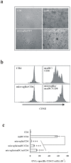
Figure 1. Microglia inhibit maDC-initiated proliferation of CD4 T cells.
CD4 T cells from DO11.10×C57BL/6 F1 hybrid mice were cocultured with maDCs or/and microglia for 5 days. (a)The photos were taken in bright field (objective ×20). (b) The proliferation of CFSE-labeled CD4 T cells was detected by FACS after 3 days coculture. (c) The live CD4 T cells in the coculture system were counted by FACS. Data are presented as mean±s.d. of triplicate wells and representative of three independent experiments. *** P<0.01.
Figure 2
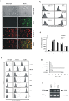
Figure 2. The characteristics of the endothelial cells from the CNS.
(a) The CNS endothelial stroma cells before and after CD11b CD31+ selection were observed under the microscopy. The purity of CD11b CD31+ cells was tested by expression of CD31 and CD106. (b, c) The secretion of cytokines including IL-7, TGF-β, GM-CSF, M-CSF and VEGF(b), and chemokines including MCP-1, MCP-3, SDF-1α, TECK and MDC(c) was detected. (d)The chemotactic ability to monocytes or DCs of the supernatant of LPS-treated EC was assayed.
Figure 3
Figure 3. Endothelia induce maDCs to differentiate into MLCs.
(a) GFP+ maDCs cultured on monolayers of CNS endothelia for 14 days and were labeled with PE-conjugated anti-CD11b antibody. Fluorescence photos were taken with a Leica fluorescent microscope (objective 40×). (b) Comparison of phenotype among microglia, MLCs and maDCs. (c) Comparison of phagocytic ability among microglia, MLCs and maDCs. Numbers in histograms indicate the geometric mean fluorescence. (d) The secretion of IL-12p70, IL-12p40, IL-10, TGF-β1 and VEGF by maDC, microglia and MLCs stimulated by 10 ng/ml LPS for 24 h was detected. (e) The production of NO by maDC, microglia and MLCs stimulated by 10 ng/ml LPS for 24 h was detected. (f) The expression of iNOS in maDC, microglia and MLCs was detected by RT-PCR.
Figure 4
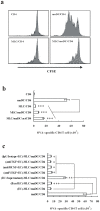
Figure 4. MLCs inhibit maDC-induced proliferation of CD4 T cells.
CD4 T cells from DO11.10×C57BL/6 F1 hybrid mice were cocultured with maDCs and/or MLCs for 5 days. (a) Proliferation of CD4 T cells in the coculture system were demonstrated by CFSE dilution. (b) MLCs were added into the maDC/CD4 T coculture system simultaneously or 24 h after maDC coculture with CD4 T cells. Live CD4 T cells in the coculture system were counted by a flow cytometer. Data are presented as mean±s.d. of triplicate wells and representative of three independent experiments. (c) Fixed endothelial cells (EC) or endothelial supernatant were used to culture DC or neutralizing antibodies against TGF-β, M-CSF or VEGF were added into the endothelial-DC coculture system(5 ug/ml, each antibody), then the differentiated MLC were added into the maDC/T cell coculture system. 3 days later, the alive T cells were counted by FACS. **, P<0.05. ***, P<0.01.
Figure 5
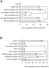
Figure 5. NO is involved in the immune inhibition by microglia and MLCs.
(a) The inhibitory function of supernatants(SN) of microglia or MLCs cultured for 48 hours and paraformaldehyde-fixed microglia or MLCs were tested in the mDC/CD4 coculture system. (b) NO donor NOC-18 at a dosage of 40 µg/ml, NO synthetase inhibitor PBIT at a dosage of 20 µg/ml and neutralization antibodies against IL-10 were added into the maDCs/T cells coculture system and then the live CD4 T cells were detected by FACS. Data are presented as mean±s.d. of triplicate wells and representative of three independent experiments.*, P>0.05;**, P<0.05; ***, P<0.01.
Figure 6
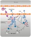
Figure 6. A model of dynamic transformation of antigen presenting cells in CNS.
Some special infection in the CNS can destroy neural tissues and endothelial cells of capillary, in this status, BBB was impaired followed by the entrance of monocytes and autoreactive T cells from peripheral blood. Monocytes will become mature DCs in the stimulation of inflammation factors secreted by the infected cells, after crossing the endothelia. New mature DCs derived from monocytes will capture the antigens of neural tissue, prime autoreactive T cells and lead to immunopathological injury. On the contrary, microglia activated by the inflammation factors can secrete high level of NO to inhibit T cell proliferation, even induce T cell apoptosis. The role of IL-10 secreted by microglia in the inhibitory function remains to be investigated. The infiltrated dendritic cells after priming might become inhibitory microglia like cells or microglia under the sustained influence of microenvironment. The cell-to-cell interaction played a key role in the differentiation of DC. The effects of TGF-β, M-CSF, VEGF secreted by activated endothelial cells remain to be investigated. Whether MLC will differentiate into microglia is unknown. Microglia and the transformation from priming dendritic cells to inhibitory microglia like cells and even microglia contribute to the remission of the autoimmune diseases of CNS.

References

    1. Hanisch UK, Kettenmann H. Microglia: active sensor and versatile effector cells in the normal and pathologic brain. Nat Neurosci. 2007;10:1387–1394. - PubMed
    1. Tambuyzer BR, Ponsaerts P, Nouwen EJ. Microglia: gatekeepers of central nervous system immunology. J Leukoc Biol. 2009;85:352–370. - PubMed
    1. Ulvestad E, Williams K, Bjerkvig R, Tiekotter K, Antel J, et al. Human microglial cells have phenotypic and functional characteristics in common with both macrophages and dendritic antigen-presenting cells. J Leukoc Biol. 1994;56:732–740. - PubMed
    1. Bailey SL, Carpentier PA, McMahon EJ, Begolka WS, Miller SD. Innate and adaptive immune responses of the central nervous system. Crit Rev Immunol. 2006;26:149–188. - PubMed
    1. Medawar PB. Immunity to homologous grafted skin; the fate of skin homografts transplanted to the brain, to subcutaneous tissue, and to the anterior chamber of the eye. Br J Exp Pathol. 1948;29:58–69. - PMC - PubMed

Publication types

Substances