. 2009 Nov 18;4(11):e7792.
doi: 10.1371/journal.pone.0007792.

SMF-1, SMF-2 and SMF-3 DMT1 orthologues regulate and are regulated differentially by manganese levels in C. elegans

Affiliations

SMF-1, SMF-2 and SMF-3 DMT1 orthologues regulate and are regulated differentially by manganese levels in C. elegans

Catherine Au et al. PLoS One. .

Abstract

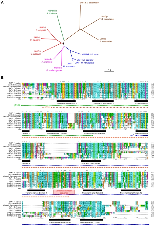
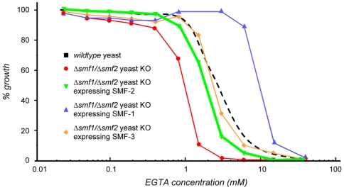
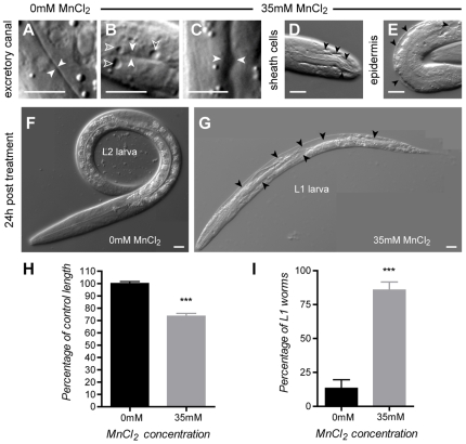
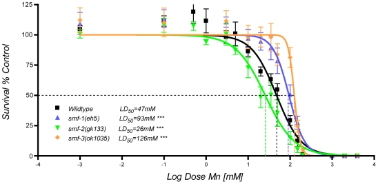
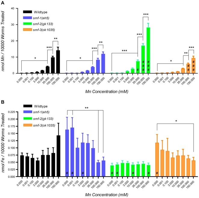
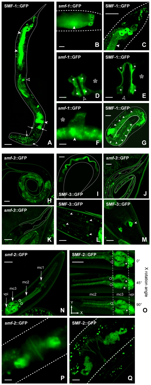
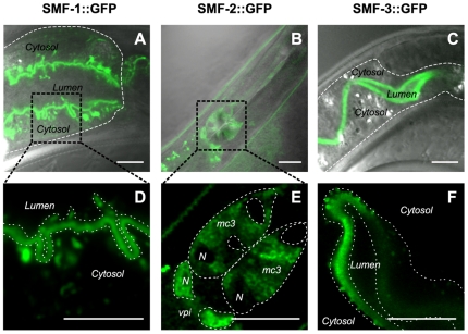
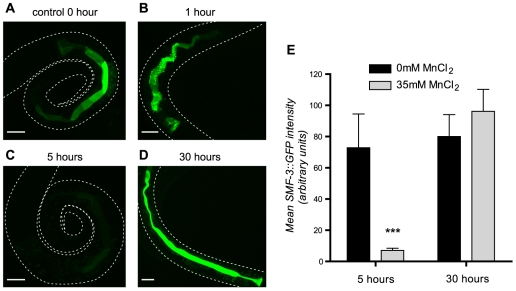
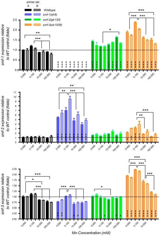
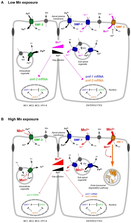
Manganese (Mn) is an essential metal that can exert toxic effects at high concentrations, eventually leading to Parkinsonism. A major transporter of Mn in mammals is the divalent-metal transporter (DMT1). We characterize here DMT1-like proteins in the nematode C. elegans, which regulate and are regulated by Mn and iron (Fe) content. We identified three new DMT1-like genes in C. elegans: smf-1, smf-2 and smf-3. All three can functionally substitute for loss of their yeast orthologues in S. cerevisiae. In the worm, deletion of smf-1 or smf-3 led to an increased Mn tolerance, while loss of smf-2 led to increased Mn sensitivity. smf mRNA levels measured by QRT-PCR were up-regulated upon low Mn and down-regulated upon high Mn exposures. Translational GFP-fusions revealed that SMF-1 and SMF-3 strongly localize to partially overlapping apical regions of the gut epithelium, suggesting a differential role for SMF-1 and SMF-3 in Mn nutritional intake. Conversely, SMF-2 was detected in the marginal pharyngeal epithelium, possibly involved in metal-sensing. Analysis of metal content upon Mn exposure in smf mutants revealed that SMF-3 is required for normal Mn uptake, while smf-1 was dispensable. Higher smf-2 mRNA levels correlated with higher Fe content, supporting a role for SMF-2 in Fe uptake. In smf-1 and smf-3 but not in smf-2 mutants, increased Mn exposure led to decreased Fe levels, suggesting that both metals compete for transport by SMF-2. Finally, SMF-3 was post-translationally and reversibly down-regulated following Mn-exposure. In sum, we unraveled a complex interplay of transcriptional and post-translational regulations of 3 DMT1-like transporters in two adjacent tissues, which regulate metal-content in C. elegans.


Conflict of interest statement

Competing Interests: The authors have declared that no competing interests exist.

Figures

Figure 1
Figure 1. The C. elegans genome encodes 3 SMF transporters orthologous to the plant, fungi and animal DMT protein family.
(A) Unrooted phylogenetic tree of a subset of eukaryotic members of the DMT family of transporters. C. elegans SMF proteins are more closely related to the animal than to the fungus or plant orthologues. (B) Multiple alignment of animal DMT1 orthologues. The 12-transmembrane domain topology of vertebrate DMT1 is conserved in C. elegans SMF proteins (black boxes), as well as the consensus transport sequence (red box). Dotted arrows indicate regions of the proteins affected by the deletion alleles eh5, gk113, and ok1035 of smf-1, 2, and 3 respectively. Amino acids with similar biochemical properties are highlighted with the same color. * represent residues conserved in all aligned sequences, : corresponds to highly conserved residues and. to less conserved residues.
Figure 2
Figure 2. C. elegans SMF transporters rescue EGTA sensitivity of yeast ΔSmf1Smf-2 mutant.
S. cerevisiae double-mutant Smf1Δ+Smf-2Δ is hypersensitive to exposure to the divalent cation chelator EGTA (red), when compared to wildtype (dotted black line). Transvections of C. elegans smf-1 (blue), smf-2 (green) or smf-3 (orange) cDNA rescue the double-mutant hypersensitivity to EGTA.
Figure 3
Figure 3. Mn exposure leads to severe osmoregulation defects and developmental delay.
(A) Excretory canal in a control wild type L1 larva (solid white arrow heads). (B, C) Enlargement of the excretory canal in L1 larvae acutely exposed to 35 mM MnCl2, after 24 h (solid white arrowheads) is associated with vacuolization (hollow arrowheads). Vacuoles are also observed in the sheath cells of the chemosensory organs (D) and in the epidermis (E). (F) Control larva 24 h after 0 mM MnCl2 treatment. (G) Dying vacuolated (black arrowheads) larva 24 h after 35 mM MnCl2 exposure. (H) worms exposed to 35 mM MnCl2 (grey) are about 30% shorter than control animals (black). I, most larvae exposed to 35 mM MnCl2 are still in L1 stage at 24 h post-treatment when control animals are L2. Error bars represent SEM, *** p<0.001, scale bars are 5 µm.
Figure 4
Figure 4. Dose-response lethality curves reveal a differential sensitivity to Mn exposure for smf mutants compared to wild type worms.
Upon 30 min exposure to MnCl2 as L1 larvae the lethal concentration 50 (LD50) at which half of the worms were dead at 24 h, was 47 mM for wildtype worms (black, N = 12), 93 mM for smf-1 mutants (blue, N = 6), 26 mM for smf-2 mutants (green, N = 7), and 126 mM for smf-3 mutants (orange, N = 5). Error bars represent SEM, *** p<0.001.
Figure 5
Figure 5. Variations in Mn and Fe content in smf mutant worms upon Mn exposure.
(A) WT and smf mutants take up Mn in a dose-dependent manner. smf-2(gk133) (green) takes up significantly more Mn than WT (black) and other mutant worms following exposure to 35 (# p<0.05), 100 (## p<0.001) and 150 mM (### p<0.001) MnCl2. smf-3(ok1035) (orange) mutants take up significantly less Mn than other worms at 100 (## p<0.01) and 150 mM (### p<0.001). (B) Fe content varies differentially in smf mutants and WT upon Mn exposure. smf-2(gk133) (green) display significantly lower Fe levels (# p<0.05), while smf-1(eh5) and smf-3(ok1035) mutants show higher Fe levels (# p<0.05) than WT in absence of Mn treatment (0 mM), and at very low Mn concentration for smf-1(eh5) (0.001 mM). Error bars represent SEM. While # designate significant differences between genetic backgrounds exposed to the same manganese dose, * indicate significant differences between exposure doses within the same C. elegans strain: #/* p<0.05. ##/** p<0.01, ###/*** p<0.001.
Figure 6
Figure 6. Expression pattern analysis of C. elegans smf genes.
A, SMF-1::GFP strongly localizes to the anterior and posterior intestine (solid white arrowheads), to the anchor cell (hollow arrowhead) and to head neurons (white arrows). smf-1::GFP and SMF-1::GFP reveal expression of smf-1 gene in rectal gland cells (B,C, black asterisks), in the uterus (uv1, uv2, utse syncytium, D,E, solid white asterisks) as well as in the adult spermatheca (F) and the L1 hyp7 epidermis (G, white arrowheads). Dotted lines outline the cuticle of the worm. Hollow asterisks indicate position of fertilized embryos. Hollow arrowheads indicate position of the vulva. smf-3 is mainly expressed in the intestine as revealed by smf-3::GFP (H) and SMF-3::GFP (I), in the major epidermis hyp7 (J, dotted line) and head epidermis hyp1-6 (K, dotted line), and in head (L) and tail neurons (M). An antero-posterior gradient of smf-2 expression is noticeable in the 9 marginal epithelial cells of the pharynx (mc1, mc2, mc3) and the 6 vpi cells of the pharyngeo-intestinal valve (N and O). Fainter expression is consistently observed in the proximal gonad (P, Q). Scale bars are 5 µm.
Figure 7
Figure 7. Subcellular localization of SMF::GFP reporters.
SMF-1::GFP mostly localizes to the apical plasma membrane of the intestine and sub-apical compartments (A, D, dotted line in A underlines the basolateral membrane of the intestine, dotted lines in D delimit its apical plasma membrane). SMF-2::GFP is seen in cytoplasmic organelles in mc and vpi cells (E, cell plasma membranes are marked by a dotted line, N indicate the position of the nuclei). SMF-3::GFP is mainly restricted to the apical plasma membrane of the intestine and apical vesicular organelles (C, F, dotted line in C underlines the basolateral membrane of the intestine, dotted lines in D delimit its apical plasma membrane). Scale bars are 5 µm.
Figure 8
Figure 8. SMF-3::GFP is down-regulated upon Mn exposure.
SMF-3::GFP signal is strongly detected at the apical plasma membrane prior Mn treatment (A). After 1 hour of exposure to 35 mM MnCl2, SMF-3-GFP localizes to sub-apical vesicular compartments (B). C, SMF-3::GFP signal is strongly decreased at 5 hours post-treatment. After a day of recovery, SMF-3::GFP expression returns to control levels and SMF-3::GFP relocates to the apical plasma membrane (D). Scale bars are 5 µm. (E) quantification of apical plasma-membrane SMF-3::GFP in the whole intestine after 5 and 30 h of exposure in control and treated animals. While # designate significant differences between genetic backgrounds exposed to the same manganese dose, * indicate significant differences between exposure doses within the same C. elegans strain: #/* p<0.05. ##/** p<0.01, ###/p<0.001.
Figure 9
Figure 9. Genotype and Mn exposure influence on smf gene expression.
Both independent primer sets corresponding to the 3′ end of the cDNA and used to assess smf gene expression (set A bright colors, and set B faded colors) give consistent results (A, B, C). smf-1 and smf-3 mRNA levels display similar variations, increasing at low Mn exposure (0.1 mM) and decreasing upon high Mn concentrations (10 mM and 100 mM) in WT, smf-1 and smf-3 mutants (A, C, black, blue, orange). smf-2 mRNA levels follow the same tendency in smf-1 and smf-3 mutants but are not affected by Mn exposure in WT (B). Independent of Mn exposure, smf-1(eh5) mutant is characterized by a strong up-regulation of smf-2 expression (B, blue). Compared to other genotypes, smf expression in smf-2(gk133) does not appear to correlate with Mn exposure (A, B, C, green). The smf-3(ok1035) mutant exhibits higher smf gene expression levels regardless of Mn exposure dose (A, B, C, orange). While # designate significant differences between genetic backgrounds exposed to the same manganese dose, * indicate significant differences between exposure doses within the same C. elegans strain: #/* p<0.05. ##/** p<0.01, ###/p<0.001. Displayed significance levels between treatments reflect the weakest significance score obtained between both primer sets.
Figure 10
Figure 10. Working model for Mn and Fe uptake by SMF transporters in C. elegans.
A: Regulation of Mn, Fe contents and SMF transporters upon low Mn exposure (0.001 mM to 3 mM), which is believed to be beneficial for the worm physiology . B: Regulation of Mn, Fe contents and SMF transporters upon high Mn exposure (50 mM to 150 mM), which was shown to be toxic (Fig. 3, 4). We propose that SMF-3* is the main transporter responsible for Mn uptake (A), and that it is degraded upon exposure to high Mn concentrations (B). Since high Fe content limit Mn uptake, SMF-3 may be inhibited by intracellular Fe (A). SMF-1 would be involved in Mn uptake to a lesser extent, and together with SMF-2*, would be responsible for Fe uptake. Upon Low Mn exposure SMF-2 would be mostly required for Fe uptake (A), whereas upon high Mn exposure, SMF-2 would be inhibited and SMF-1 would partially compensate for Fe uptake (B). In the case of SMF-2 and SMF-1, metal uptake could essentially take place in acidified endosomal compartments, as SMF-2 is mainly cytoplasmic and SMF-1 is detected in sub-apical compartments. Gap-junction communications between pharyngeal epithelia, vpi cells and intestinal cells permit Mn2+ and Fe2+ to diffuse distant from their site of uptake, allowing metal-dependent regulation of smf mRNA stability or transcription. A prediction of our model is that the Fe gradient established by SMF-2 activity would be reversed upon high Mn exposure (B), and could constitute the signal for smf expression regulation. Since basal smf mRNA levels depend on the integrity of each smf genomic sequence (Fig. 9), transcriptions of smf genes are assumed to be interdependent, maybe because they require a common transcription factor. * SMF-2 might transport Mn and SMF-3 might transport Fe, but these possibilities are not explored in this model. The size of the text reflects the concentration of the species.

References

    1. Post JE. Manganese oxide minerals: crystal structures and economic and environmental significance. Proc Natl Acad Sci U S A. 1999;96:3447–3454. - PMC - PubMed
    1. Saric M. Bronchial hyperreactivity and occupational asthma. Am J Ind Med. 1986;9:217–219. - PubMed
    1. Wedler FC, Denman RB. Glutamine synthetase: the major Mn(II) enzyme in mammalian brain. Curr Top Cell Regul. 1984;24:153–169. - PubMed
    1. Addess KJ, Basilion JP, Klausner RD, Rouault TA, Pardi A. Structure and dynamics of the iron responsive element RNA: implications for binding of the RNA by iron regulatory binding proteins. J Mol Biol. 1997;274:72–83. - PubMed
    1. Aschner M, Gannon M, Kimelberg HK. Manganese uptake and efflux in cultured rat astrocytes. J Neurochem. 1992;58:730–735. - PubMed

Publication types

Substances