. 2009 Nov 16;4(11):e7774.
doi: 10.1371/journal.pone.0007774.

Transcriptional regulation and biological functions of selenium-binding protein 1 in colorectal cancer in vitro and in nude mouse xenografts

Affiliations

Transcriptional regulation and biological functions of selenium-binding protein 1 in colorectal cancer in vitro and in nude mouse xenografts

Nicole M Pohl et al. PLoS One. .

Abstract

Background: It has been shown that selenium-binding protein 1 (SBP1) is significantly downregulated in different human cancers. Its regulation and function have not yet been established.

Methodology and principal findings: We show that the SBP1 promoter is hypermethylated in colon cancer tissues and human colon cancer cells. Treatment with 5'-Aza-2'-deoxycytidine leads to demethylation of the SBP1 promoter and to an increase of SBP1 promoter activity, rescues SBP1 mRNA and protein expression in human colon cancer cells. Additionally, overexpression of SBP1 sensitizes colon cancer cells to H2O2-induced apoptosis, inhibits cancer cell migration in vitro and inhibits tumor growth in nude mice.

Conclusion and significance: These data demonstrate that SBP1 has tumor suppressor functions that are inhibited in colorectal cancer through epigenetic silencing.


Conflict of interest statement

Competing Interests: The authors have declared that no competing interests exist.

Figures

Figure 1
Figure 1. SBP1 promoter was hypermethylated in human cancer tissues and colon cancer cell lines, which silenced gene expression and was revered by 5-Aza-2′-Deoxycytidine (DAC).
A, SBP1 promoter was highly methylated in human colon cancer tissues. Genomic DNA from adjacent normal (N) and adenocarcinoma (T) tissue was isolated, converted and amplified for human SBP1 promoter using MS-PCR. # indicated different patients. Unmethylated (U) and methylated (M) bands were detected by DNA separation on 2% agarose gels with ethidium bromide. B, Western blotting analysis showed various expression of SBP1 in human colon cancer cell lines; HCT116 cells with stable transfection of SBP1 overexpressing plasmid was used as positive control. C, SBP1 promoter was hypermethylated in HCT116 cells. Genomic DNA from LS174T, HCT116, SW480, Caco-2 and HT-29 colon cancer cells was isolated, converted and amplified via MS-PCR for human SBP1 promoter followed by gel separation of total DNA on 2% agarose gels (U, unmethylated DNA; M, methylated DNA). D, DAC reversed SBP1 promoter methylation in HCT116 cells. DNA from HCT116 cells treated with 30 µM of DAC for 96 h was isolated and separated on 2% agarose gels after conversion and MS-PCR. E, Luciferase assay showed that DAC increased SBP1 promoter luciferase activity of HCT116 cells with transfection of a vector containing the full length promoter of SBP1 and with a treatment of 30 µM DAC or PBS for 72 hours. pGL4 vector and Renilla was co-transfected as controls (* p<0.01, compared to PBS). F, DAC restored SBP1 protein expression in HCT116 cells with DAC treatment for 72 h, analyzed by Western blotting. G. SBP1 mRNA was restored by DAC in HCT116 cells (* p<0.01, compared to PBS). Each experiment was performed at least 3 times.
Figure 2
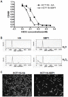
Figure 2. SBP1 had tumor suppressive functions.
A, HCT116 cells transfected with HA-SBP1 had an increased sensitivity to H2O2 by MTS proliferation assay compared to the empty vector (HA). The cells were treated with H2O2 for 24 hours. B, HCT116 cells transfected with HA-SBP1 increased H2O2-induced apoptosis by Flow cytometry analysis. Cells were treated with 0.3 mM of H2O2 for 24 hours. Boxes indicate apoptotic cells (sub-G1). C, SBP1 inhibited cancer cell migration: cell migration of SBP1 overexpressing HCT116 cells and HA-control cells was assessed via transwell chambers. Migrated cells were detected with DAPI staining.
Figure 3
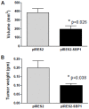
Figure 3. BP1 attenuated colorectal cancer cell growth in NIH-III nude mice.
NIH-III nude mice were injected with HCT116 cells stably expressing SBP1 (pIRES2-SBP1) or vector (pIRES2) control. Four weeks after injection, tumor volume (A) and tumor weight (B) was determined.

References

    1. Chang PW, Tsui SK, Liew C, Lee CC, Waye MM, et al. Isolation, characterization, and chromosomal mapping of a novel cDNA clone encoding human selenium binding protein. J Cell Biochem. 1997;64:217–224. - PubMed
    1. Porat A, Sagiv Y, Elazar Z. A 56-kDa Selenium-binding Protein Participates in Intra-Golgi Protein Transport. J Biol Chem. 2000;275:14457–14465. - PubMed
    1. Scortegagna M, Martin RJ, Kladney RD, Neumann RG, Arbeit JM. Hypoxia-Inducible Factor-1{alpha} Suppresses Squamous Carcinogenic Progression and Epithelial-Mesenchymal Transition. Cancer Research. 2009;69:2638–2646. - PMC - PubMed
    1. Jeong J-Y, Wang Y, Sytkowski AJ. Human selenium binding protein-1 (hSP56) interacts with VDU1 in a selenium-dependent manner. Biochemical and Biophysical Research Communications. 2009;379:583–588. - PubMed
    1. Yang M, Sytkowski AJ. Differential Expression and Androgen Regulation of the Human Selenium-binding Protein Gene hSP56 in Prostate Cancer Cells. Cancer Research. 1998;58:3150–3153. - PubMed

Publication types

Substances