. 2010 May;28(3):276-88.
doi: 10.1007/s00774-009-0134-y. Epub 2009 Nov 19.

Overexpression of heme oxygenase-1 increases human osteoblast stem cell differentiation

Affiliations

Overexpression of heme oxygenase-1 increases human osteoblast stem cell differentiation

Ignazio Barbagallo et al. J Bone Miner Metab. 2010 May.

Abstract

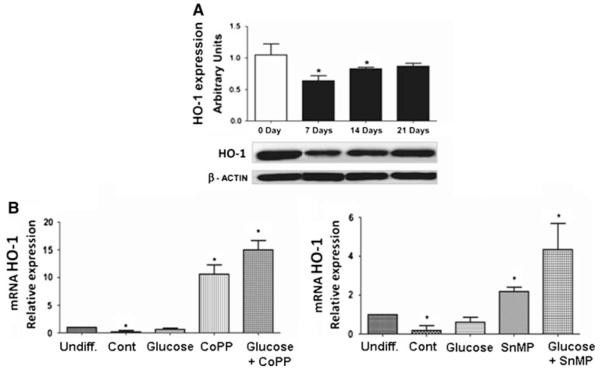
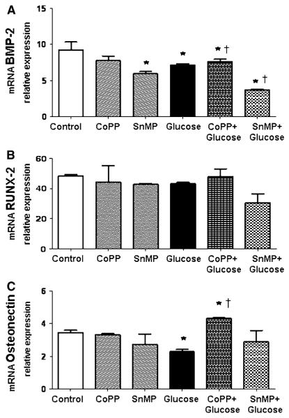
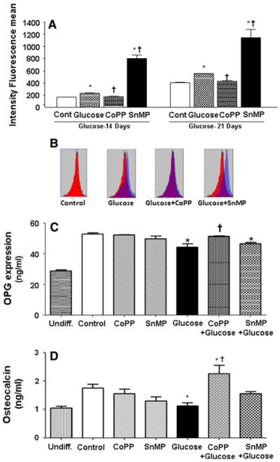
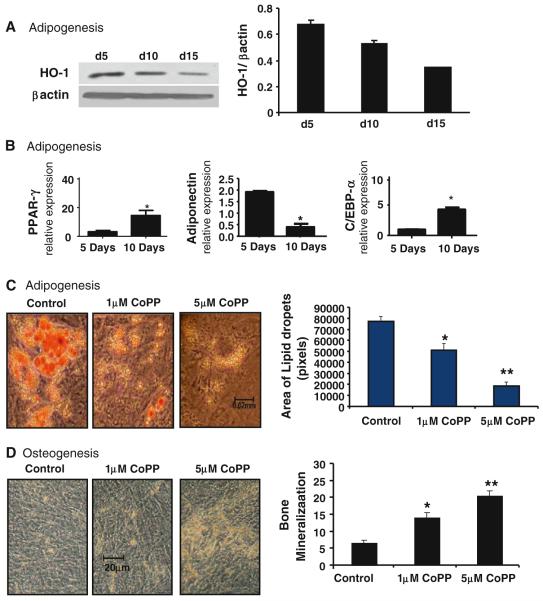
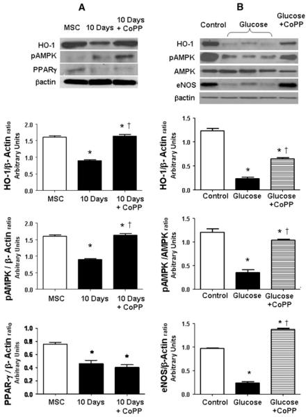
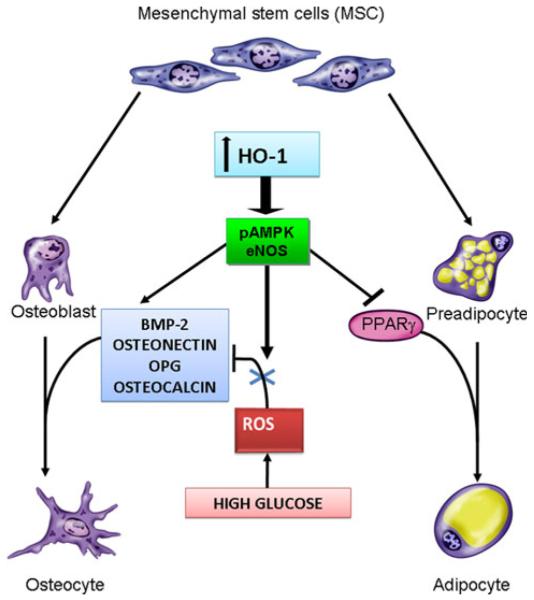
Human bone marrow mesenchymal stem cells (MSCs) are pleiotrophic cells that differentiate to either adipocytes or osteoblasts as a result of crosstalk by specific signaling pathways including heme oxygenase (HO)-1/-2 expression. We examined the effect of inducers of HO-1 expression and inhibitors of HO activity on MSC differentiation to the osteoblast and following high glucose exposure. MSC cultured in osteogenic medium increased expression of osteonectin, Runt-related transcription factor 2 (RUNX-2), osteocalcin, and alkaline phosphatase. HO-1 expression during differentiation was initially decreased and then followed by a rebound increase after 15 days of culture. Additionally, the effect of HO-1 on osteoblasts appears different to that seen in adipocyte stem cells. On addition of a cobalt compound, the resultant induction of HO-1 decreases adipogenesis. Moreover, glucose (30 mM) inhibited osteoblast differentiation, as evidenced by decreased bone morphogenetic protein (BMP)-2, osteonectin, osteocalcin, and osteoprotegerin (OPG). In contrast, MSC-derived adipocytes were increased by glucose. Increased HO-1 expression increased the levels of osteonectin, OPG, and BMP-2. Inhibition of HO activity prevented the increase in osteonectin and potentiated the decrease of osteocalcin and OPG in cells exposed to high glucose levels. Furthermore, targeting HO-1 expression increased pAMPK and endothelial nitric oxide synthase (eNOS) and restored osteoblastic markers. Our findings suggest that targeting HO-1 gene expression attenuates the hyperglycemia-mediated decrease in MSC-derived osteoblast differentiation. Finally, the mechanism underlying the HO-1-specific cell effect on osteoblasts and adipocytes is yet to be explored. Thus, the targeting of HO-1 gene expression presents a portal to increase osteoblast function and differentiation and attenuate osteoporosis by promoting bone formation.


Figures

Fig. 1
Fig. 1
Osteoblast differentiation and relative markers. a–f Morphological features of undifferentiated and osteoblastic differentiated mesenchymal stem cells (MSCs). Light microscopy analysis of cells after 14 days (b) and 21 days (c) of culture in osteogenic differentiation media shows a gradual increase in the number of osteoblastic cells compared with undifferentiated MSCs (a). d–f Presence of mineralized nodules following alizarin red staining analysis of undifferentiated cells (d) and at 14 (e) and 21 (f) days after culture in osteogenic differentiation media. g–k Expression of osteoblastic markers in differentiated MSCs by quantitative real-time polymerase chain reaction (qRT-PCR) and enzyme-linked immunosorbent assay (ELISA) testing. qRT-PCR reveals a marked increase in bone morphogenetic protein (BMP)-2 (g), Runt-related transcription factor 2 (RUNX-2) (h), and osteonectin (i) at 21 days of osteoblastic differentiation. ELISA shows an increase of osteocalcin secreted in the medium at 21 days with respect to undifferentiated cells (j). k Alkaline phosphatase (AP) activity during osteoblastic differentiation from BM MSCs. Bars represent the mean ± SEM of three independent experiments. *P < 0.05 versus undifferentiated cells; n = 4
Fig. 2
Fig. 2
a Development pattern of heme oxygenase (HO)-1 mRNA and protein expression in undifferentiated cells, and at 7, 14, and 21 days. Shown are changes in HO-1 mRNA (upper panel), analyzed by qRT-PCR, and protein expression (lower panel), analyzed by Western blot during osteoblastic differentiation of MSCs. The profile of HO-1 mRNA and protein expression during osteoblastic differentiation shows that HO-1 is significantly downregulated during osteoblastic differentiation. b Effect of CoPP, HO-1 inducers (left panel), and HO inhibitor (SnMP) (right panel) on HO-1 mRNA levels after 21 days of osteoblastic differentiation. Bars represent the mean ± SEM of three independent experiments. *P < 0.05 versus undifferentiated cells (hMSCs); n = 3
Fig. 3
Fig. 3
Effect of CoPP 0.5 μM, SnMP 5 μM, and glucose 30 mM on BMP-2 (a), RUNX-2 (b), and osteonectin (c) after 21 days of osteoblastic differentiation. Bars represent the mean ± SEM of four independent experiments. *P < 0.05 versus osteogenic medium (OM); P < 0.05 versus glucose 30 mM (OM + glucose); n = 5
Fig. 4
Fig. 4
a Quantification of reactive oxygen species (ROS) by cytofluorimetric analysis during osteoblastic differentiation. Osteoblastic differentiation resulted in a slight ROS production starting from 14 days of differentiation. b ROS emission spectrum obtained after 21 days of osteoblastic differentiation. c Osteoprotegerin (OPG) secretion during differentiation measured in the medium by ELISA. CoPP 0.5 μM treatment in presence of high glucose concentration (OM + glucose + CoPP) is able to restore the OPG level to similar values of OM and osteocalcin secretion. d During differentiation, measured in the medium by ELISA, CoPP 0.5 μM (OM + glucose + CoPP) treatment at high glucose levels, osteoblastic differentiation was able to induce osteocalcin secretion with respect to exposure to high glucose only. CoPP treatment in the presence of glucose 30 mM (OM + glucose + CoPP) shows a decrease of mRNA expression with respect to OM + glucose. Bars represent the mean ± SEM of four independent experiments. *P < 0.05 versus osteogenic medium (OM); P < 0.05 versus glucose 30 mM (OM + glucose)
Fig. 5
Fig. 5
Effect of increased HO-1 on adipogenesis and osteogenesis in hMSC. a Basal levels of HO-1 in adipogenesis of hMSC on days 5, 10, and 15 were measured by Western blot. b mRNA expression levels of PPAR-γ, adiponectin, and C/EBPα in adipogenesis-treated hMSCs for days 5 and 10, respectively. c hMSCs were also treated with 1 and 5 μM CoPP in adipogenesis media for 14 days. Area and size of lipid droplets were determined by measuring individual lipid droplets (pixel area) from three different fields using ImagePro software. d hMSCs were also treated with 1 and 5 μM CoPP in osteogenesis media for 14 days, and osteocytes were stained with alizarin red. Levels of significance: *P < 0.05, **P < 0.01 control versus CoPP; n = 4
Fig. 6
Fig. 6
a Western blots and densitometer analysis of HO-1, pAMPK, PPAR-γ, and β-actin in undifferentiated cells (MSC) and at 10 days of osteoblast differentiation in presence or absence of CoPP treatment. Bars represent the mean ± SEM of four independent experiments. *P < 0.05 versus undifferentiated cells (MSC); P < 0.05 versus 10 days. b Western blots and densitometer analysis of HO-1, pAMPK, AMPK, eNOS, and β-actin in differentiated cells (control), in high glucose differentiated cells (glucose), and after CoPP treatment in high glucose osteoblast differentiation. Bars represent the mean ± SEM of four independent experiments. *P < 0.05 versus differentiated cells (control); P < 0.05 versus glucose; n = 3
Fig. 7
Fig. 7
Scheme of the mechanisms of HO-1 regulation of osteoblast and adipocyte differentiation. Hyperglycemia causes an increase in ROS, resulting in the overproduction of superoxide and subsequent impairment in osteoblast differentiation by a decrease of BMP-2, osteonectin, osteocalcin, and OPG. Targeting HO-1 by a cobalt compound increased the levels of osteonectin, OPG, and BMP-2. Furthermore, targeting HO-1 expression increased pAMPK and eNOS, reduced ROS formation, and restored osteoblastic markers, suggesting an antiinflammatory or antioxidant role

References

    1. Ferrari G, Cusella-De AG, Coletta M, Paolucci E, Stornaiuolo A, Cossu G, Mavilio F. Muscle regeneration by bone marrow-derived myogenic progenitors. Science. 1998;279:1528–1530. - PubMed
    1. Pittenger MF, Mackay AM, Beck SC, Jaiswal RK, Douglas R, Mosca JD, Moorman MA, Simonetti DW, Craig S, Marshak DR. Multilineage potential of adult human mesenchymal stem cells. Science. 1999;284:143–147. - PubMed
    1. Marie PJ, Fromigue O. Osteogenic differentiation of human marrow-derived mesenchymal stem cells. Regen Med. 2006;1:539–548. - PubMed
    1. Barbagallo I, Tibullo D, Di RM, Giallongo C, Palumbo GA, Raciti G, Campisi A, Vanella A, Green CJ, Motterlini R. A cytoprotective role for the heme oxygenase-1/CO pathway during neural differentiation of human mesenchymal stem cells. J Neurosci Res. 2008;86:1927–1935. - PubMed
    1. Hamilton EJ, Rakic V, Davis WA, Chubb SA, Kamber N, Prince RL, Davis TM. Prevalence and predictors of osteopenia and osteoporosis in adults with type 1 diabetes. Diabet Med. 2009;26:45–52. - PubMed

Publication types

MeSH terms