. 2010 Nov;22(11):2491-502.
doi: 10.1162/jocn.2009.21384.

Hemicraniectomy: a new model for human electrophysiology with high spatio-temporal resolution

Affiliations

Hemicraniectomy: a new model for human electrophysiology with high spatio-temporal resolution

Bradley Voytek et al. J Cogn Neurosci. 2010 Nov.

Abstract

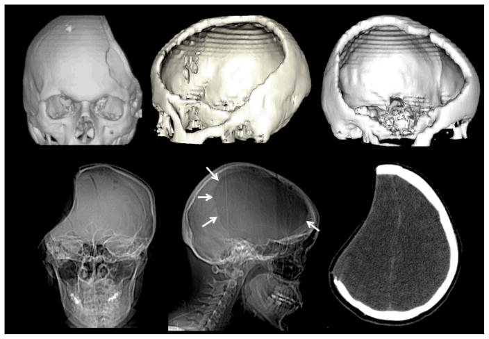
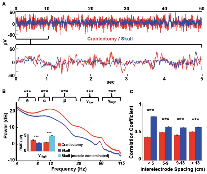
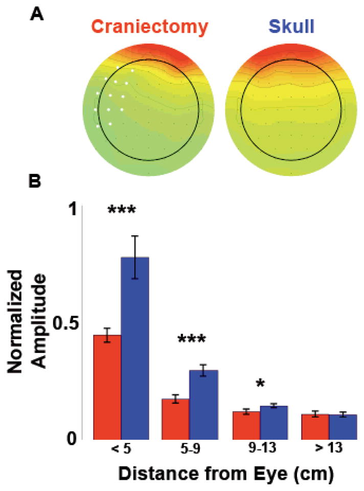
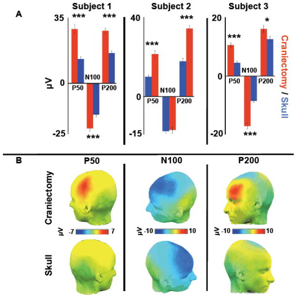
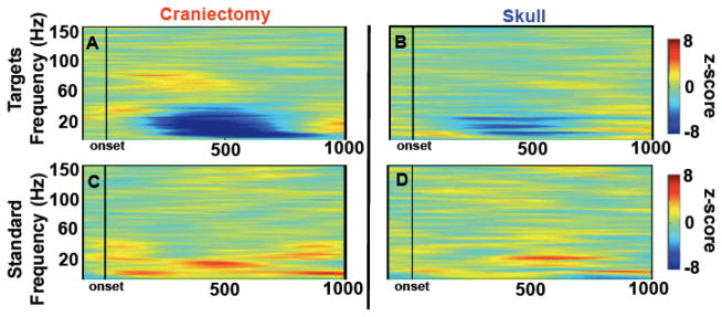
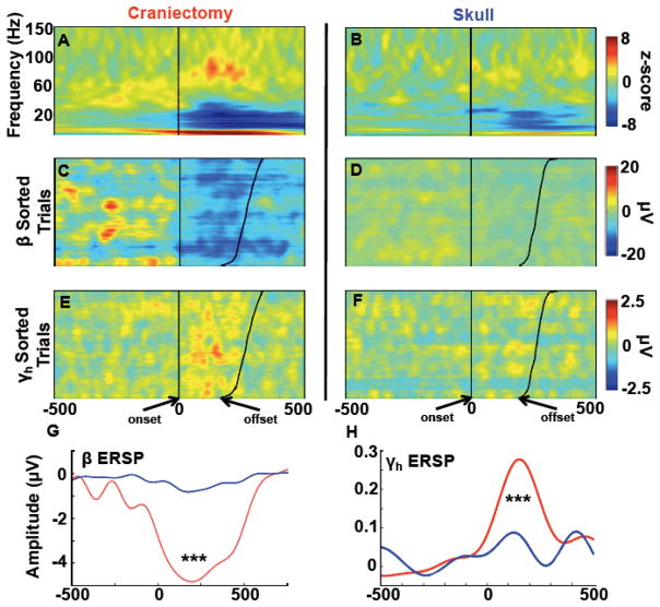
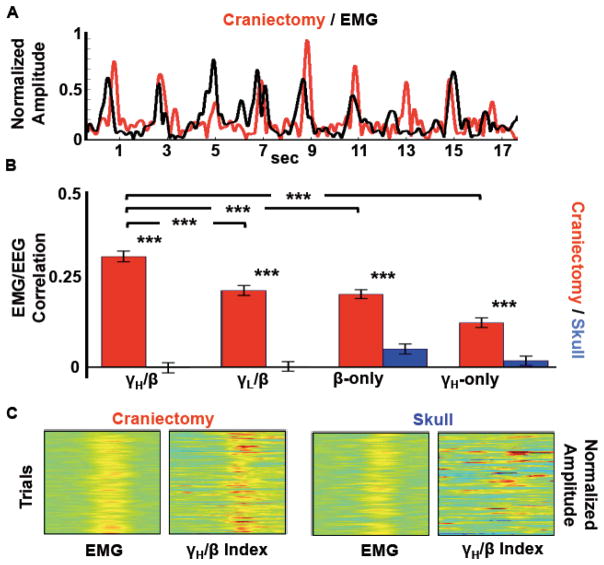
Human electrophysiological research is generally restricted to scalp EEG, magneto-encephalography, and intracranial electrophysiology. Here we examine a unique patient cohort that has undergone decompressive hemicraniectomy, a surgical procedure wherein a portion of the calvaria is removed for several months during which time the scalp overlies the brain without intervening bone. We quantify the differences in signals between electrodes over areas with no underlying skull and scalp EEG electrodes over the intact skull in the same subjects. Signals over the hemicraniectomy have enhanced amplitude and greater task-related power at higher frequencies (60-115 Hz) compared with signals over skull. We also provide evidence of a metric for trial-by-trial EMG/EEG coupling that is effective over the hemicraniectomy but not intact skull at frequencies >60 Hz. Taken together, these results provide evidence that the hemicraniectomy model provides a means for studying neural dynamics in humans with enhanced spatial and temporal resolution.


Figures

Figure 1
Figure 1
Computed tomography (CT) scans of the three subjects demonstrating the extent of the hemicraniectomy. The upper row images were reconstructed using “Surface 3D volume-rendered” multidetector helical CT data. The lower row images include AP and lateral plain radiographs and an axial CT image. Note how the calvarial defect (arrows) includes virtually the entire the frontal bone with relative sparing of the supraorbital rim and paramedian skull. It extends into the parietal bone and squamosal portion of the temporal bone down to just above the ear. The axial CT image demonstrates the characteristic concave deformity seen in many patients prior to cranioplasty. Note the immediate proximity of the skin surface to the underlying normal brain.
Figure 2
Figure 2
Comparison of raw signals. A, Time-series data has more power and decreased signal redundancy over the hemicraniectomy (red) as compared to the skull (blue). The top trace shows 50 seconds of raw data from two homologous electrodes from subject 1 (F3/F4); the bottom is zoomed in on the first 5 seconds of the top trace. B, Mean power spectra for hemicraniectomy and skull electrodes not contaminated by muscle noise (width of spectrum indicates s.e.m. for each frequency; not 60 Hz contamination). Asterisks above each band grouping indicate root-mean-square (RMS) amplitude for that frequency band is greater for hemicraniectomy electrodes compared to homologous skull electrodes. For γH (65–115 Hz) RMS analyses electrodes were divided into two groups: skull electrodes with high muscle noise and those with low (inset; see Supplemental Methods for muscle noise classification methods). γH is greater over hemicraniectomy sites compared to skull electrodes only when skull electrodes are not contaminated by muscle noise. C, Interelectrode correlations for all hemicraniectomy pairs are lower at all distances compared to homologous skull pairs. ***p < 0.001, statistically significant differences; bars are s.e.m.
Figure 3
Figure 3
Blink artifact propagation. A, Scalp topographies of mean peri-blink amplitude for all three hemicraniectomy subjects compared to three young controls with intact skull, demonstrating the blink amplitude drop over the hemicraniectomy electrodes. Electrodes over the site of hemicraniectomy in any one subject are in white. B, Peri-blink amplitudes as a function of distance from the eye. ***p < 0.001, *p = 0.042, statistically significant differences; bars are s.e.m.
Figure 4
Figure 4
Auditory ERPs in response to correctly identified infrequent deviant tones. A, Peak ERP amplitudes for components P50, N100, and P200 over hemicraniectomy and homologous skull sites for each subject. B, Grand average scalp topographies of mean amplitude across the time range of each ERP for all three subjects. ***p < 0.001, *p = 0.033, statistically significant differences; bars are s.e.m.
Figure 5
Figure 5
Stimulus-locked auditory time-frequency activity. A, B, Event-related spectral perturbations for subjects 1 and 3 over homologous hemicraniectomy and skull sites showing target-related γH over the hemicraniectomy. C, D, Same as A and B to non-target auditory stimuli. Note the early cluster of γH starting at approximately 100 ms post-stimulus onset is only present over the hemicraniectomy in response to targets.
Figure 6
Figure 6
Contralateral movement-related time-frequency activity. (Note that figures AF are on the same x-axis scale). A, B, Event-related spectral perturbations for all three subjects over homologous hemicraniectomy and skull sites showing movement-related γH over the hemicraniectomy. C, D, Single trial β (12–30 Hz) and E, F, γH (65–115 Hz) movement-related activity in subject 1 locked to movement onset—sorted by movement offset—illustrating the trial-by-trial power at hemicraniectomy electrode (C3) compared to the homologous skull electrode (C4). G, H, β and γH ERPs for all subjects. Movement-related β and γH are greater over hemicraniectomy electrodes as compared to homologous skull sites. ***p < 0.001, statistically significant differences; bars are s.e.m.
Figure 7
Figure 7
EEG/EMG movement correlation. A, Example of raw traces of EMG and contralateral γH/β index in subject 1 over hemicraniectomy electrode C3. B, Average trial-by-trial EMG/EEG correlations for all subjects. For all correlation bands, hemicraniectomy electrodes perform better than skull electrodes. For hemicraniectomy electrodes, γH/β correlates higher with movement than γL/β, β-only, and γH-only. C, Single-trial movement-locked EMG and contralateral γH/β index in subject 1 over hemicraniectomy (C3) and skull (C4) illustrating trial-by-trial correlation between γH/β index and EMG. ***p < 0.001, statistically significant differences; bars are s.e.m.

References

    1. Abraham K, Marsan C. Patterns of cortical discharges and their relation to routine scalp electroencephalography. Electroencephalography and Clinical Neurophysiology. 1958;10(3):447–461. - PubMed
    1. Ball T, Demandt E, Mutschler I, Neitzel E, Mehring C, Vogt K, Aertsen A, Schulzebonhage A. Movement related activity in the high gamma range of the human EEG. NeuroImage. 2008;41(2):302–310. - PubMed
    1. Ball T, Kern M, Mutschler I, Aertsen A, Schulze-Bonhage A. Signal quality of simultaneously recorded invasive and non-invasive EEG. NeuroImage. 2009;46 (3):708–716. - PubMed
    1. Bédard C, Kröger H, Destexhe A. Does the 1/f frequency scaling of brain signals reflect self-organized critical states? Physical Review Letters. 2006;97(11):11802-1–4. - PubMed
    1. Bénar CG, Gotman J. Modeling of post-surgical brain and skull defects in the EEG inverse problem with the boundary element method. Clinical Neurophysiology. 2002;113(1):48–56. - PubMed

Publication types

MeSH terms