

Metabolic levels in the corpus callosum and their structural and behavioral correlates after moderate to severe pediatric TBI
- PMID: 19925210
- PMCID: PMC2867590
- DOI: 10.1089/neu.2009.1058
Metabolic levels in the corpus callosum and their structural and behavioral correlates after moderate to severe pediatric TBI
Abstract
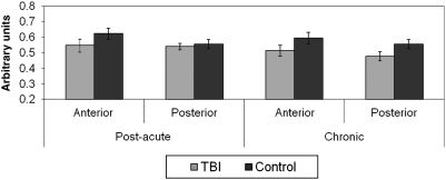
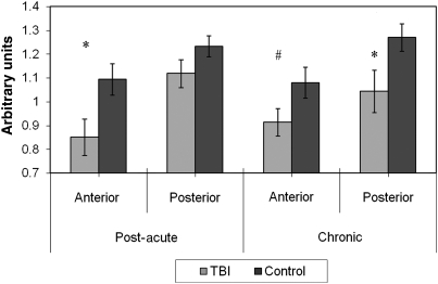
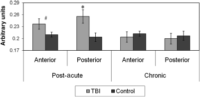
Diffuse axonal injury (DAI) secondary to traumatic brain injury (TBI) contributes to long-term functional morbidity. The corpus callosum (CC) is particularly vulnerable to this type of injury. Magnetic resonance spectroscopy (MRS) was used to characterize the metabolic status of two CC regions of interest (ROIs) (anterior and posterior), and their structural (diffusion tensor imaging; DTI) and neurobehavioral (neurocognitive functioning, bimanual coordination, and interhemispheric transfer time [IHTT]) correlates. Two groups of moderate/severe TBI patients (ages 12-18 years) were studied: post-acute (5 months post-injury; n = 10), and chronic (14.7 months post-injury; n = 8), in addition to 10 age-matched healthy controls. Creatine (energy metabolism) did not differ between groups across both ROIs and time points. In the TBI group, choline (membrane degeneration/inflammation) was elevated for both ROIs at the post-acute but not chronic period. N-acetyl aspartate (NAA) (neuronal/axonal integrity) was reduced initially for both ROIs, with partial normalization at the chronic time point. Posterior, not anterior, NAA was positively correlated with DTI fractional anisotropy (FA) (r = 0.88), and most domains of neurocognition (r range 0.22-0.65), and negatively correlated with IHTT (r = -0.89). Inverse corerlations were noted between creatine and posterior FA (r = -0.76), neurocognition (r range -0.22 to -0.71), and IHTT (r = 0.76). Multimodal studies at distinct time points in specific brain structures are necessary to delineate the course of the degenerative and reparative processes following TBI, which allows for preliminary hypotheses about the nature and course of the neural mechanisms of subsequent functional morbidity. This will help guide the future development of targeted therapeutic agents.
Figures
References
-
- Akpinar E. Koroglu M. Ptak T. Diffusion tensor MR imaging in pediatric head trauma. J. Comput. Assist. Tomogr. 2007;31:657–661. - PubMed
-
- Ashwal S. Babikian T. Gardner-Nichols J. Freier M.C. Tong K.A. Holshouser B.A. Susceptibility-weighted imaging and proton magnetic resonance spectroscopy in assessment of outcome after pediatric traumatic brain injury. Arch. Phys. Med. Rehabil. 2006a;87:S50–S58. - PubMed
-
- Ashwal S. Holshouser B.A. Shu S.K. Simmons P.L. Perkin R.M. Tomasi L.G. Knierim D.S. Sheridan C. Craig K. Andrews G.H. Hinshaw D.B. Predictive value of proton magnetic resonance spectroscopy in pediatric closed head injury. Pediatr. Neurol. 2000;23:114–125. - PubMed
-
- Ashwal S. Holshouser B.A. Tong K.A. Use of advanced neuroimaging techniques in the evaluation of pediatric traumatic brain injury. Dev. Neurosci. 2006b;28:309–326. - PubMed
Publication types
MeSH terms
Substances
Grants and funding
LinkOut - more resources
Full Text Sources
