

The expression of Gli3, regulated by HOXD13, may play a role in idiopathic congenital talipes equinovarus
- PMID: 19925654
- PMCID: PMC2784749
- DOI: 10.1186/1471-2474-10-142
The expression of Gli3, regulated by HOXD13, may play a role in idiopathic congenital talipes equinovarus
Abstract
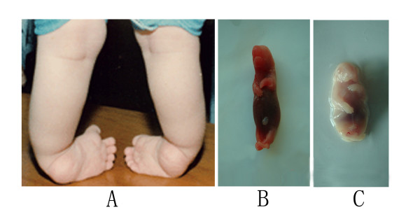
Background: Idiopathic congenital talipes equinovarus (ICTEV) is a congenital limb deformity. Based on extended transmission disequilibrium testing, Gli-Kruppel family member 3 (Gli3) has been identified as a candidate gene for ICTEV. Here, we verify the role of Gli3 in ICTEV development.
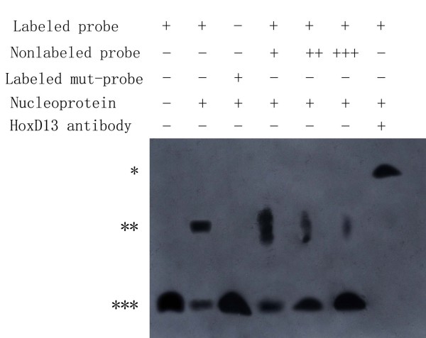
Methods: Using the rat ICTEV model, we analyzed the differences in Gli3 expression levels between model rats and normal control rats. We used luciferase reporter gene assays and ChIP/EMSA assays to analyze the regulatory elements of Gli3.
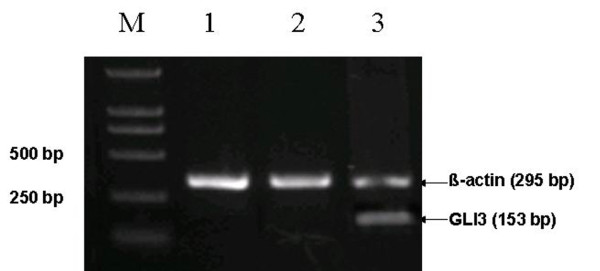
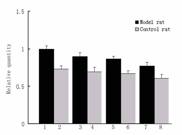
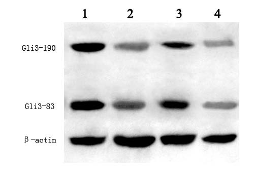
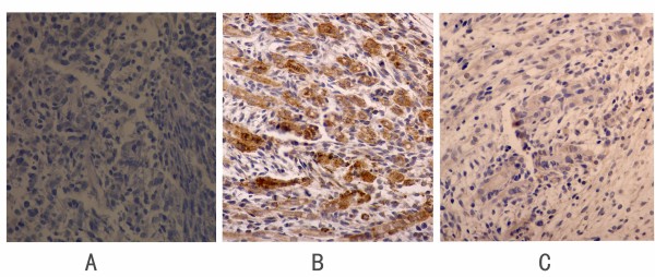
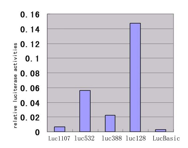
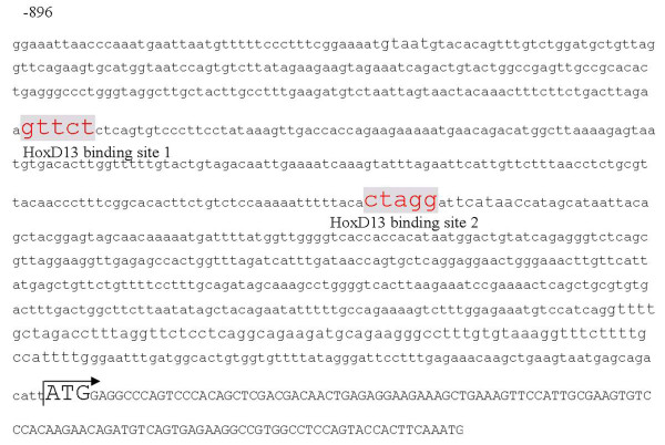
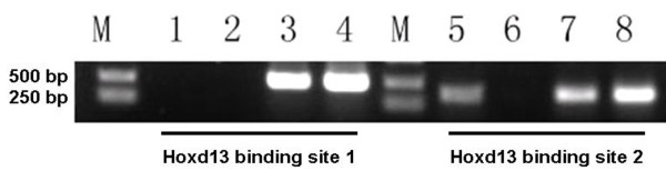
Results: Gli3 showed higher expression levels in ICTEV model rats compared to controls (P < 0.05). We identified repressor and activator regions in the rat Gli3 promoter. The Gli3 promoter also contains two putative Hoxd13 binding sites. Using EMSA, the Hoxd13 binding site 2 was found to directly interact with Hoxd13 in vitro. ChIP assays of the Hoxd13-Gli3 promoter complex from a developing limb confirmed that endogenous Hoxd13 interacts with this region in vivo.
Conclusion: Our findings suggest that HoxD13 directly interacts with the promoter of Gli3. The increase of Gli3 expression in ICTEV model animal might result from the low expression of HoxD13.
Figures
Similar articles
-
The etiology of idiopathic congenital talipes equinovarus: a systematic review.J Orthop Surg Res. 2018 Aug 22;13(1):206. doi: 10.1186/s13018-018-0913-z. J Orthop Surg Res. 2018. PMID: 30134936 Free PMC article.
-
[Mechanism of GLI3 gene transcription regulation in idiopathic congenital talipes equinovarus].Zhonghua Yi Xue Yi Chuan Xue Za Zhi. 2012 Oct;29(5):537-41. doi: 10.3760/cma.j.issn.1003-9406.2012.05.008. Zhonghua Yi Xue Yi Chuan Xue Za Zhi. 2012. PMID: 23042389 Chinese.
-
[The functions of GLI3 and EN1 genes in idiopathic congenital talipes equinovarus].Yi Chuan. 2009 Dec;31(12):1214-20. doi: 10.3724/sp.j.1005.2009.01214. Yi Chuan. 2009. PMID: 20042388 Chinese.
-
[Possible role of GLI3 gene in the pathogenesis of idiopathic congenital talipes equinovarus].Zhonghua Yi Xue Yi Chuan Xue Za Zhi. 2012 Jun;29(3):260-5. doi: 10.3760/cma.j.issn.1003-9406.2012.03.003. Zhonghua Yi Xue Yi Chuan Xue Za Zhi. 2012. PMID: 22678783 Chinese.
-
A systematic review of association studies of common variants associated with idiopathic congenital talipes equinovarus (ICTEV) in humans in the past 30 years.Springerplus. 2016 Jun 27;5(1):896. doi: 10.1186/s40064-016-2353-8. eCollection 2016. Springerplus. 2016. PMID: 27386344 Free PMC article. Review.
Cited by
-
Apoptotic genes expression in placenta of clubfoot-like fetus pregnant rats.Int J Clin Exp Pathol. 2014 Jan 15;7(2):677-84. eCollection 2014. Int J Clin Exp Pathol. 2014. PMID: 24551289 Free PMC article.
-
Integrated bioinformatics analysis of potential pathway biomarkers using abnormal proteins in clubfoot.PeerJ. 2020 Jan 20;8:e8422. doi: 10.7717/peerj.8422. eCollection 2020. PeerJ. 2020. PMID: 31998564 Free PMC article.
-
Association between vascular endothelial growth factor promoter polymorphisms and the risk of recurrent implantation failure.Exp Ther Med. 2018 Feb;15(2):2109-2119. doi: 10.3892/etm.2017.5641. Epub 2017 Dec 15. Exp Ther Med. 2018. PMID: 29434813 Free PMC article.
-
The etiology of idiopathic congenital talipes equinovarus: a systematic review.J Orthop Surg Res. 2018 Aug 22;13(1):206. doi: 10.1186/s13018-018-0913-z. J Orthop Surg Res. 2018. PMID: 30134936 Free PMC article.
-
Gli3 regulation of myogenesis is necessary for ischemia-induced angiogenesis.Circ Res. 2013 Oct 25;113(10):1148-58. doi: 10.1161/CIRCRESAHA.113.301546. Epub 2013 Sep 17. Circ Res. 2013. PMID: 24044950 Free PMC article.
References
Publication types
MeSH terms
Substances
LinkOut - more resources
Full Text Sources
Molecular Biology Databases
