. 2010 Feb;1797(2):285-95.
doi: 10.1016/j.bbabio.2009.11.005. Epub 2009 Nov 24.

Regulation of vascular smooth muscle cell bioenergetic function by protein glutathiolation

Affiliations

Regulation of vascular smooth muscle cell bioenergetic function by protein glutathiolation

Bradford G Hill et al. Biochim Biophys Acta. 2010 Feb.

Abstract

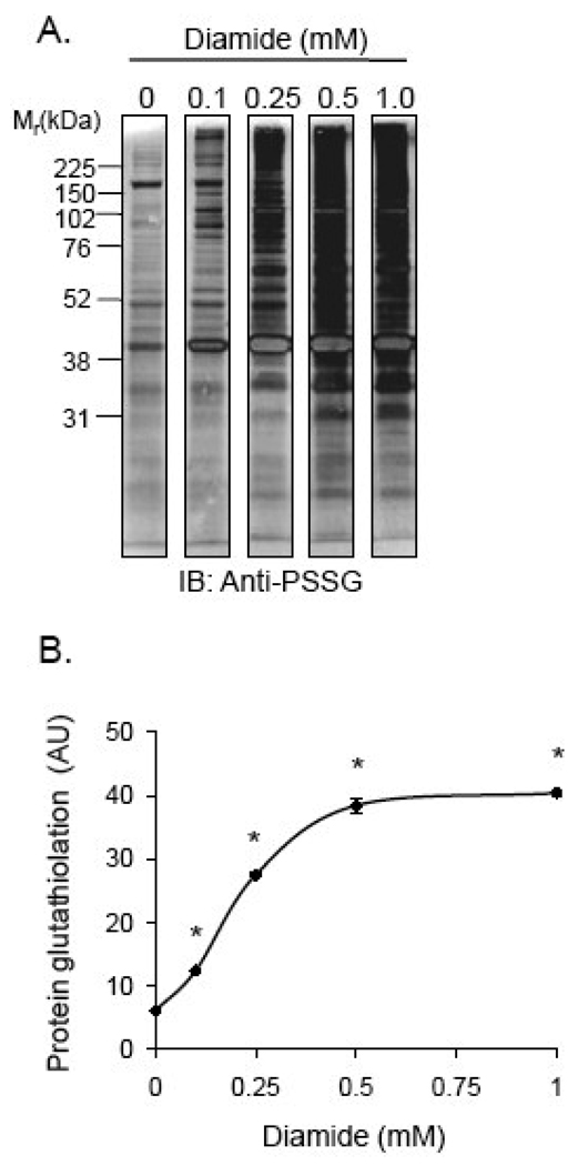
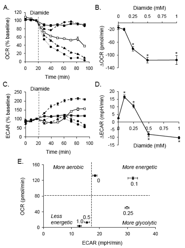
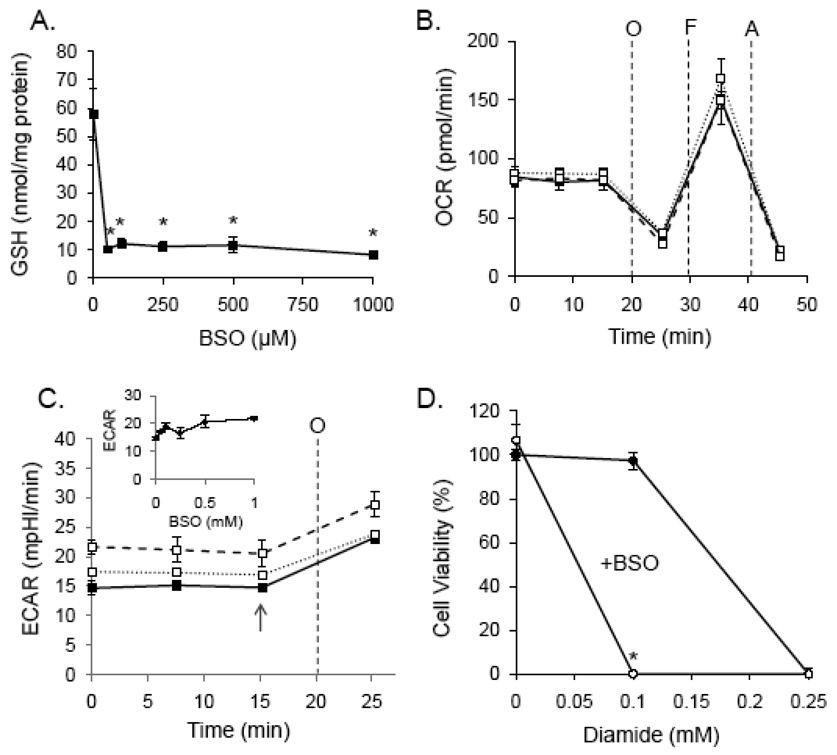
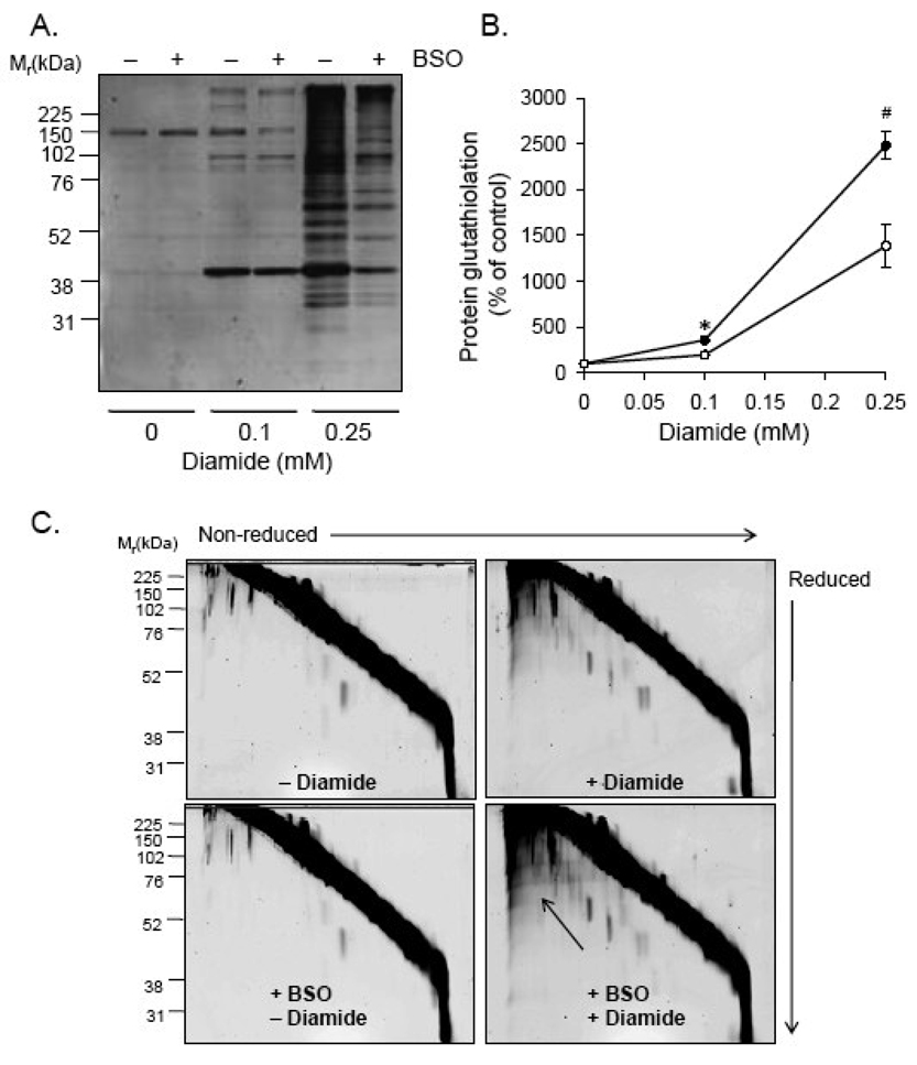
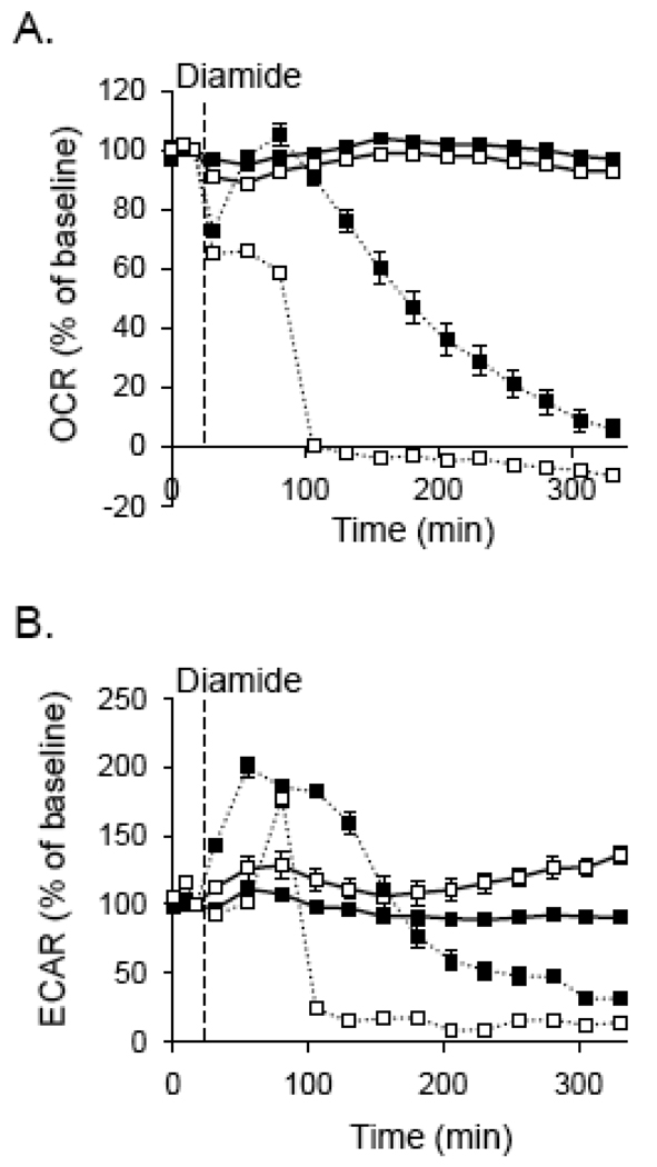
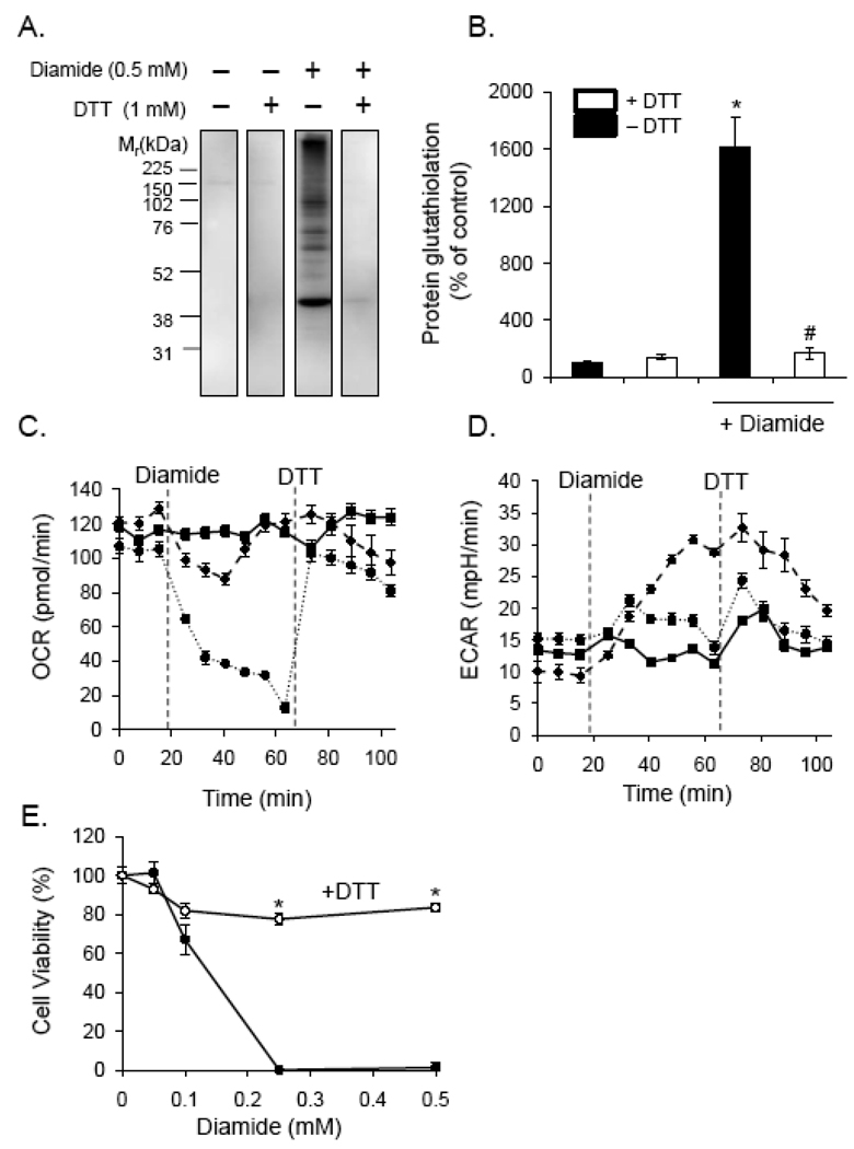
Protein thiolation by glutathione is a reversible and regulated post-translational modification that is increased in response to oxidants and nitric oxide. Because many mitochondrial enzymes contain critical thiol residues, it has been hypothesized that thiolation reactions regulate cell metabolism and survival. However, it has been difficult to differentiate the biological effects due to protein thiolation from other oxidative protein modifications. In this study, we used diamide to titrate protein glutathiolation and examined its impact on glycolysis, mitochondrial function, and cell death in rat aortic smooth muscle cells. Treatment of cells with diamide increased protein glutathiolation in a concentration-dependent manner and had comparably little effect on protein-protein disulfide formation. Diamide increased mitochondrial proton leak and decreased ATP-linked mitochondrial oxygen consumption and cellular bioenergetic reserve capacity. Concentrations of diamide above 200 microM promoted acute bioenergetic failure and caused cell death, whereas lower concentrations of diamide led to a prolonged increase in glycolytic flux and were not associated with loss of cell viability. Depletion of glutathione using buthionine sulfoximine had no effect on basal protein thiolation or cellular bioenergetics but decreased diamide-induced protein glutathiolation and sensitized the cells to bioenergetic dysfunction and death. The effects of diamide on cell metabolism and viability were fully reversible upon addition of dithiothreitol. These data suggest that protein thiolation modulates key metabolic processes in both the mitochondria and cytosol.


Figures

Fig. 1
Fig. 1. Protein glutathiolation by diamide
Analysis of protein-glutathione (PSSG) adduct formation by Western blotting: (A) Representative Western blot of PSSG adducts: Cells were treated with diamide (0–1 mM) for 1 h. Cell lysate proteins were then separated by SDS-PAGE followed by chemifluorescent Western blotting using anti-protein-glutathione (anti-PSSG) antibodies. (B) Group data from panel A. n = 3 per group; *p<0.05 vs. cells not treated with diamide.
Fig. 2
Fig. 2. Bioenergetic changes induced by protein thiolation
Extracellular flux analyses of diamide treated cells: Baseline measurements of oxygen consumption rate (OCR; panel A) and extracellular acidification rate (ECAR; panel C) were measured. Diamide was then injected where indicated to 0 (closed squares), 0.1 (diamonds; dashed line), 0.25 (open squares), 0.5 (triangles; dashed line), or 1.0 (circles; dotted line) mM final concentrations. Shown in panels A and C are the OCR and ECAR expressed as a percentage of baseline. (B and D) The changes in OCR (panel B) and ECAR (panel D) at the last time point measured are shown as a function of diamide concentration. N = 4 per group, *p < 0.05 vs. cells not treated with diamide. (E) Metabolic profile of diamide-treated cells: The last time point of OCR and ECAR measurements in panels A and C, respectively, were used to construct a 2D plot of bioenergetic function. The numbers above each point indicate the concentration of diamide (in mM).
Fig. 3
Fig. 3. Characterization of mitochondrial dysfunction induced by protein thiolation
Delineation of changes in ATP-linked oxygen consumption, proton leak, and respiratory capacity in cells treated with diamide. (A) Extracellular flux analysis was used to measure mitochondrial function in intact, adherent smooth muscle cells. To probe individual components of respiration that contributed to the consumption of oxygen, oligomycin (1 µg/ml), FCCP (1 µM), and antimycin A (10 µM) were injected sequentially. This allowed for estimation of the contribution of proton leak (Leak) and ATP demand (ATP) to mitochondrial oxygen consumption. The maximal respiratory capacity was determined using the FCCP-stimulated rate. The residual oxygen consumption that occurred after addition of antimycin A was ascribed to non-mitochondrial sources. (B) After baseline measurements of oxygen consumption rate (OCR), diamide was injected to 0 (closed squares), 0.1 (diamonds; dotted line), 0.25 (open squares), or 0.5 (triangles; dotted line) mM final concentrations. At the indicated time, oligomycin (O; 1 µg/ml) was injected and OCR was measured. FCCP (F; 1 µM) was then injected followed by another OCR measurement. Last, antimycin A (A; 10 µM) was injected and OCR was again measured. (C and D) The oligomycin-sensitive and –insensitive rates of oxygen consumption were used to calculate the amount of oxygen consumption that was linked to ATP production (panel C) and proton leak (panel D). (E) The reserve or spare respiratory capacity was calculated in control and diamide-treated cells by subtracting the maximal (FCCP-stimulated) rate of oxygen consumption from the basal OCR. N = 5 per group, *p < 0.05 vs. cells not treated with diamide.
Fig. 4
Fig. 4. Glutathione depletion does not affect mitochondrial function but potentiates diamide-induced cell death
(A) Cells were treated with buthionine sulfoximine (BSO; 0 – 1 mM) for 24 h and free glutathioine was measured by glutathione recycling assay. N ≥ 3 per group, *p < 0.05 vs. cells not treated with BSO. (B) Mitochondrial function in glutathione-depleted cells: Cells were treated with 0 (closed squares), 0.05 (open squares; dashed line), or 0.5 (open squares; dotted line) mM BSO for 24 h followed by mitochondrial function assay. After three baseline measurements of oxygen consumption rate (OCR), oligomyin (O; 1 µg/ml), FCCP (F; 1 µM), and antimycin A (A; 10 µM) were injected sequentially, and rates of oxygen consumption were recorded after each injection. (C) Glycolytic flux in glutathione-depleted cells: After 24 h of 0 (closed squares), 0.05 (open squares; dashed line), or 0.5 (open squares; dotted line) mM BSO treatment, baseline and oligomycin-stimulated extracellular acidification rates (ECAR) were measured. Inset: At the point shown by the arrow, the rate of extracellular was plotted for cells treated with 0–1 mM BSO. (D) Viability of normal (closed circles) and glutathione-depleted (open circles) cells treated with 0–0.25 mM diamide. N ≥ 3, and *p < 0.05 vs. diamide-treated cells not treated with BSO.
Fig. 5
Fig. 5. Glutathione depletion decreases diamide-induced protein-glutathione adduct formation
Protein-glutathione adducts and protein-protein dithiol formation in glutathione-depleted cells treated with diamide. (A) Protein-glutathione adducts in cells depleted of glutathione: Cells were treated with 100 µM BSO for 24 h followed by diamide (0–0.25 mM) for 40 min. Equal amounts of protein (10 µg) from cell lysates were separated by SDS-PAGE, and glutathiolated proteins were detected by chemifluorescent Western blotting with anti-PSSG antibodies. (B) Group data from panel A. n = 3 per group; *p < 0.05 vs. glutathione-depleted cells treated with 0.1 mM diamide; #p<0.05 vs. glutathione-depleted cells treated with 0.25 mM diamide. (C) Diagonal electrophoresis gels in control cells (upper left panel), cells treated with 0.25 mM diamide (upper right panel), glutathione-depleted cells (lower left panel), and glutathione-depleted cells treated with 0.25 mM diamide. The arrow points to the area of the gel showing increased protein-protein dithiol formation.
Fig. 6
Fig. 6. Bioenergetic function in glutathione-depleted cells treated with diamide
Extracellular flux analysis plots of oxygen consumption and extracellular acidification: (A) Cells were treated without or with 100 µM BSO for 24 h (open squares), and baseline oxygen consumption rates (OCR) were recorded. Diamide (dotted lines) was then injected to 0.1 mM final concentrations where indicated, and the OCR were followed for approximately 300 min. (B) Glycolytic flux in glutathione-depleted cells treated with diamide: Cells were treated as described in panel A and rates of extracellular acidification were measured. N = 5 per group.
Fig. 7
Fig. 7. De-thiolation promotes bioenergetic recovery and prevents cell death
Protein-glutathione adducts, bioenergetics, and cell viability in diamide-treated cells exposed to the reducing agent, dithiothreitol. (A) Dithiothreitol (DTT) reverses protein glutathiolation induced by diamide: Cells were treated without or with diamide for 40 min. DTT was then added to the cells to a final concentration of 1 mM for 10 min. The medium was then removed, and the cells were lysed in buffer containing N-ethylmaleimide. Protein-glutathione (PSSG) adducts were assessed by chemiluminescent Western blotting with anti-PSSG antibodies. (B) Group data from panel A. n = 3 per group; *p<0.05 vs. cells not treated with diamide; #p<0.05 vs. diamide-treated cells not exposed to DTT. (C and D) Bioenergetic measurements after diamide and DTT addition to cells: After measurement of the baseline oxygen consumption (panel C) and extracellular acidification (panel D) rates (OCR and ECAR, respectively), diamide was injected to 0 (closed squares), 0.1 (triangles; dashed line), or 0.5 (circles; dotted line) mM concentrations. At the indicated time, DTT was injected to a final concentration of 1 mM, and OCR and ECAR measurements were recorded. N = 5 per group. (E) Cell viability in diamide and DTT-treated cells. Cells were treated with diamide in the absence or presence of 1 mM DTT for 16 h. Cell viability was then measured by MTT assay. N = 4 per group; *p < 0.05 vs. cells treated with diamide alone.

References

    1. Diotte NM, Xiong Y, Gao J, Chua BH, Ho YS. Attenuation of doxorubicin-induced cardiac injury by mitochondrial glutaredoxin 2. Biochim. Biophys. Acta. 2009;1793:427–438. - PubMed
    1. Adachi T, Weisbrod RM, Pimentel DR, Ying J, Sharov VS, Schoneich C, Cohen RA. S-Glutathiolation by peroxynitrite activates SERCA during arterial relaxation by nitric oxide. Nat. Med. 2004;10:1200–1207. - PubMed
    1. Ghezzi P, Romines B, Fratelli M, Eberini I, Gianazza E, Casagrande S, Laragione T, Mengozzi M, Herzenberg LA, Herzenberg LA. Protein glutathionylation: coupling and uncoupling of glutathione to protein thiol groups in lymphocytes under oxidative stress and HIV infection. Mol. Immunol. 2002;38:773–780. - PubMed
    1. Eaton P, Byers HL, Leeds N, Ward MA, Shattock MJ. Detection, quantitation, purification, and identification of cardiac proteins S-thiolated during ischemia and reperfusion. J. Biol. Chem. 2002;277:9806–9811. - PubMed
    1. Lind C, Gerdes R, Schuppe-Koistinen I, Cotgreave IA. Studies on the mechanism of oxidative modification of human glyceraldehyde-3-phosphate dehydrogenase by glutathione: catalysis by glutaredoxin. Biochem. Biophys. Res. Commun. 1998;247:481–486. - PubMed

Publication types

MeSH terms

LinkOut - more resources