. 2010 Feb 12;396(1):105-17.
doi: 10.1016/j.jmb.2009.11.019. Epub 2009 Nov 17.

Organization, structure, and assembly of alpha-carboxysomes determined by electron cryotomography of intact cells

Affiliations

Organization, structure, and assembly of alpha-carboxysomes determined by electron cryotomography of intact cells

Cristina V Iancu et al. J Mol Biol. .

Abstract

Carboxysomes are polyhedral inclusion bodies that play a key role in autotrophic metabolism in many bacteria. Using electron cryotomography, we examined carboxysomes in their native states within intact cells of three chemolithoautotrophic bacteria. We found that carboxysomes generally cluster into distinct groups within the cytoplasm, often in the immediate vicinity of polyphosphate granules, and a regular lattice of density frequently connects granules to nearby carboxysomes. Small granular bodies were also seen within carboxysomes. These observations suggest a functional relationship between carboxysomes and polyphosphate granules. Carboxysomes exhibited greater size, shape, and compositional variability in cells than in purified preparations. Finally, we observed carboxysomes in various stages of assembly, as well as filamentous structures that we attribute to misassembled shell protein. Surprisingly, no more than one partial carboxysome was ever observed per cell. Based on these observations, we propose a model for carboxysome assembly in which the shell and the internal RuBisCO (ribulose-1,5-bisphosphate carboxylase/oxygenase) lattice form simultaneously, likely guided by specific interactions between shell proteins and RuBisCOs.


Figures

Figure 1
Figure 1
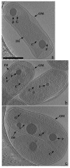
Overview of the three organisms examined: a) H. neapolitanus; b) T. intermedia; c) T. crunogena. A wealth of structural features can be seen including high-density granules (P), carboxysomes (C), internal granules and fibers (G), and filamentous structures (F). Also visible are the inner (IM) and outer membranes (OM). All images representing cells here and throughout are tomographic slices (usually 12-nm thick). Scale bar is 500 nm.
Figure 2
Figure 2
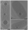
Organization of carboxysomes in H. neapolitanus cells: a) group of carboxysomes clustered together near the center of a cell; b) carboxysomes packed around an electron-dense granule; c) carboxysomes dispersed throughout the cell. Scale bar is 500 nm.
Figure 3
Figure 3
Elemental analysis of dense granules. The figure shows three images of the same H. neapolitanus cell: a) zero-loss 2-D projection image; b) 2-D projection electron energy loss spectrographic image focused on phosphorous; c) slice through the corresponding tomogram. The black arrows in a) and c) highlight dense granules; the black arrows in b) highlight regions rich in phosphorous. The white arrow in c) highlights an internal granule too small to be detected by the elemental map. Scale bars are 500 nm.
Figure 4
Figure 4
Associations of carboxysomes with polyP granules: a) H. neapolitanus cell, showing close association between carboxysomes and granules (note the string of density between the topmost granule and the adjacent carboxysomes, as well as the densities within the carboxysomes, which resemble the densities of the polyP granules); b) H. neapolitanus cell, showing indentation of the polyP granule surface where it abuts a carboxysome (note the line of density between the indented face of the granule and the adjacent carboxysome); c) H. neapolitanus cell, showing faceted granule abutting carboxysome (note the lattice emanating from this facet). Scale bars are 200 nm.
Figure 5
Figure 5
Lattices and strings: a) lattice protruding from a polyP granule and toward a neighboring carboxysome in an H. neapolitanus cell; b) lattice emanating out of the curved surface of a polyP granule and toward two adjacent carboxysomes in a T. intermedia cell; c) string between an indented polyP granule and the neighboring carboxysome; d) string connecting a polyP granule to an adjacent carboxysome (note the indentation in the polyP granule and the apparent gap in the carboxysome surface where the string is attached). Scale bar is 100 nm.
Figure 6
Figure 6
Dense granules and fibers within carboxysomes. Panels 1-4 show increasing sizes of internal granules in H. neapolitanus carboxysomes. Panels 5-8 show T. intermedia carboxysomes and panels 9-12 show T. crunogena carboxysomes. Panels 11 and 12 illustrate the dense fibers observed inside carboxysomes of T. crunogena. Scale bar is 50 nm.
Figure 7
Figure 7
Size and shape variability of cellular carboxysomes: a)-d) irregular carboxysomes; e) two adjacent icosahedral carboxysomes of different sizes. Panels a), b), c), and e) are from cells of H. neapolitanus. Panel d) is from a cell of T. intermedia. Scale bar is 50 nm.
Figure 8
Figure 8
Carboxysomes purified from H. neapolitanus: a) zero tilt image; b) 4.6 nm slice through tomographic reconstruction. Black arrows indicate different sizes of carboxysomes. Scale bar is 200 nm.
Figure 9
Figure 9
Partial carboxysomes. The figure is organized such that more complete carboxysomes are at the bottom. Note the variability of partial carboxysome shapes, ranging from sharp vertices (e.g. panels 2, 3, 6, 12) to smooth, rounded surfaces (e.g. panels 4, 7, 9, 10), and that putative RuBisCOs pack within the interior as the shell forms. Scale bar is 50 nm.
Figure 10
Figure 10
Filament-like structures: a) H. neapolitanus cell showing a twisted filament associated with carboxysomes; b) parallel bundle in H. neapolitanus; c) parallel bundle in T. intermedia; d) parallel bundle in H. neapolitanus (inset shows periodicity measurement); e) parallel bundle in H. neapolitanus near the inner membrane. Note the linear arrangement of possible RuBisCOs between the bundle and the membrane. Scale bar is 200 nm.

References

    1. Cheng S, Liu Y, Crowley CS, Yeates TO, Bobik TA. Bacterial microcompartments: their properties and paradoxes. Bioessays. 2008;30:1084–95. - PMC - PubMed
    1. Yeates T, Kerfeld C, Heinhorst S, Cannon GC, Shively JM. Protein-based organelles in bacteria: carboxysomes and related microcompartments. Nat Rev Microbiol. 2008;6:681–691. - PubMed
    1. Badger MR, Price GD. CO2 concentrating mechanisms in cyanobacteria: molecular components, their diversity and evolution. Journal of Experimental Botany. 2003;54:609–622. - PubMed
    1. Spreitzer RJ, Salvucci ME. Rubisco: structure, regulatory interactions, and possibilities for a better enzyme. Annual review of plant biology. 2002;53:449–75. - PubMed
    1. So AK, Espie GS, Williams EB, Shively JM, Heinhorst S, Cannon GC. A novel evolutionary lineage of carbonic anhydrase (epsilon class) is a component of the carboxysome shell. Journal of Bacteriology. 2004;186:623–30. - PMC - PubMed

Publication types

LinkOut - more resources