Review
. 2009 Dec;19(6):715-23.
doi: 10.1016/j.sbi.2009.10.010. Epub 2009 Nov 18.

Structures of RNA polymerase-antibiotic complexes

Affiliations
Review

Structures of RNA polymerase-antibiotic complexes

Mary X Ho et al. Curr Opin Struct Biol. 2009 Dec.

Abstract

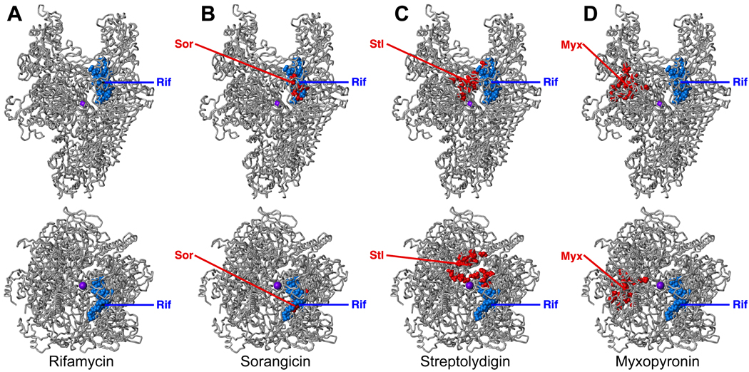
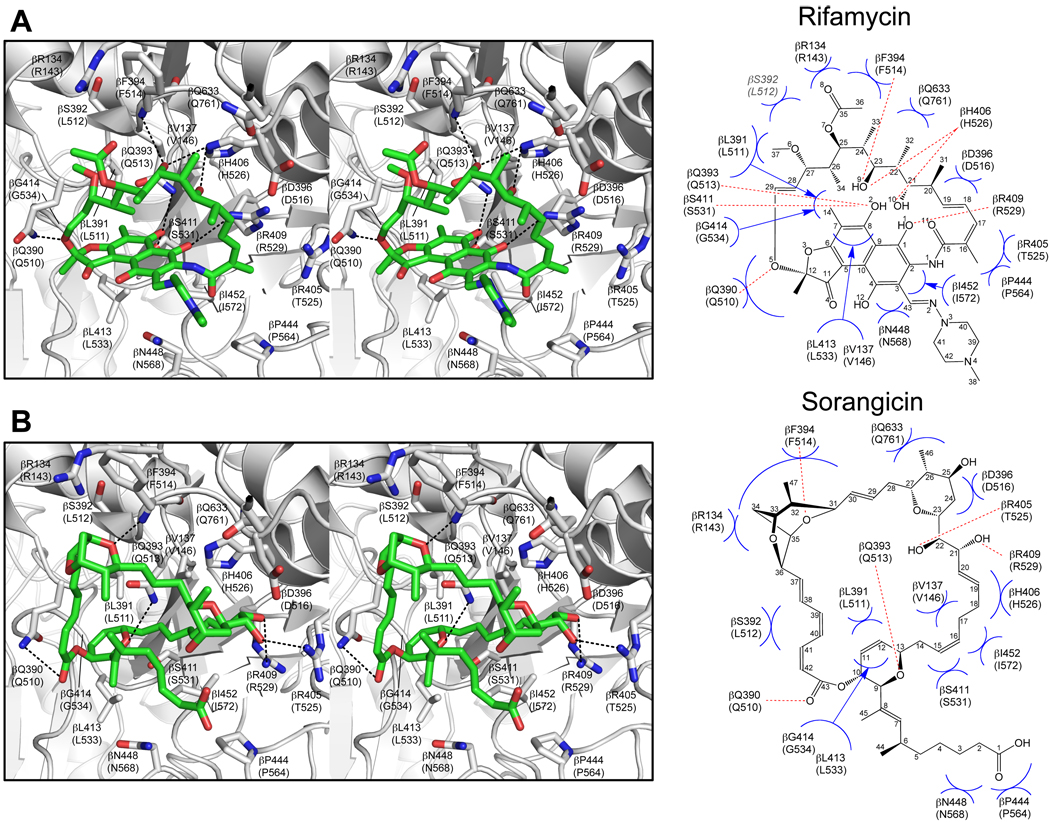
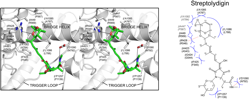
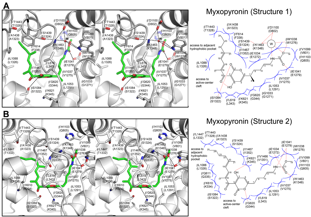
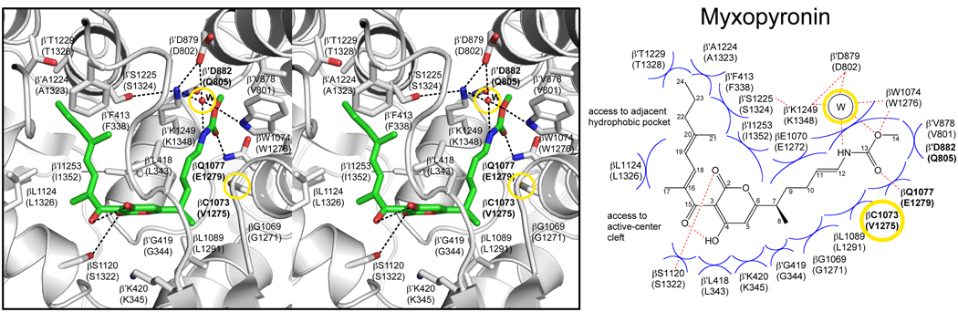
Inhibition of bacterial RNA polymerase (RNAP) is an established strategy for antituberculosis therapy and broad-spectrum antibacterial therapy. Crystal structures of RNAP-inhibitor complexes are available for four classes of antibiotics: rifamycins, sorangicin, streptolydigin, and myxopyronin. The structures define three different targets, and three different mechanisms, for inhibition of bacterial RNAP: (1) rifamycins and sorangicin bind near the RNAP active center and block extension of RNA products; (2) streptolydigin interacts with a target that overlaps the RNAP active center and inhibits conformational cycling of the RNAP active center; and (3) myxopyronin interacts with a target remote from the RNAP active center and functions by interfering with opening of the RNAP active-center cleft to permit entry and unwinding of DNA and/or by interfering with interactions between RNAP and the DNA template strand. The structures enable construction of homology models of pathogen RNAP-antibiotic complexes, enable in silico screening for new antibacterial agents, and enable rational design of improved antibacterial agents.


Figures

Figure 1
Figure 1. Targets of small-molecule inhibitors of RNAP
(A) Target of rifamycins (Rif; [,;–16]). (B) Target of sorangicin (Sor; [,; E. Sineva and R.H.E., unpublished]). (C) Target of streptolydigin (Stl; [–32]). (D) Target of myxopyronin (Myx; [39,40]). Each panel shows two orthogonal views of RNAP. Red, sites of substitutions conferring resistance to the specified inhibitor; blue, sites of substitutions conferring resistance to rifamycins; violet sphere, active-center Mg2+.
Figure 2
Figure 2. Structural basis of transcription inhibition by rifamycins and sorangicin: "Rif/Sor target."
(A) Crystal structure of T. aquaticus RNAP-rifampin complex (PDB 1YNN; [6]). (B) Crystal structure of T. aquaticus RNAP-sorangicin complex (PDB 1YNJ; [20]). Left subpanels: RNAP-inhibitor contacts (stereoview). Gray, RNAP backbone (ribbon representation) and RNAP sidechain carbon atoms (stick representation); green, rifampin carbon atoms; red, oxygen atoms; blue, nitrogen atoms. Dashed lines, H-bonds. Right subpanels: schematic summary of RNAP-inhibitor contacts. Red dashed lines, H-bonds. Blue arcs, van der Waals interactions. RNAP residues are numbered both as in the crystal structure of Thermus sp. RNAP and, in parentheses, as in E. coli RNAP.
Figure 3
Figure 3. Structural basis of transcription inhibition by streptolydigin: "bridge-helix/trigger-loop target."
Crystal structure of T. thermophilus RNAP-streptolydigin complex (PDB 1ZYR [30]; see also [31,32]). Left and right subpanels and residue numbering as in Figure 2.
Figure 4
Figure 4. Structural basis of transcription inhibition by myxopyronin: "switch-region target."
(A) Crystal structure of complex of T. thermophilus RNAP and myxopyronin A (PDB 3DXJ [39]). (B) Crystal structure of complex of T. thermophilus RNAP and 8-desmethyl-myxopyronin B (PDB 3EQL [40]). As compared to myxopyronin A, 8-desmethyl-myxopyronin B lacks a methyl group at the start of its enecarbamate sidechain (right sidechain) and contains one additional carbon atom at the end of its dienone sidechain (left sidechain). Left and right subpanels and residue numbering as in Figure 2. “W,” in panel (A), interfacial water molecule.
Figure 5
Figure 5. Homology model of the M. tuberculosis RNAP-Myx complex
Left and right subpanels as in Figure 2. Bold labels, RNAP residues that differ in the homology model of the M. tuberculosis complex vs. in the crystal structure of the T. thermophilus complex. “W,” interfacial water molecule present in the homology model of the M. tuberculosis complex and in the crystal structure of the T. thermophilus complex. RNAP residues are numbered both as in M. tuberculosis RNAP and, in parentheses, as in E. coli RNAP. Structural features that potentially can be exploited for design and synthesis of more potent inhibitors of M. tuberculosis RNAP are highlighted in yellow: i.e., the binding-site cysteine and the binding-site interfacial water molecule.

Similar articles

Cited by

References

    1. Chopra I. Bacterial RNA polymerase: a promising target for the discovery of new antimicrobial agents. Curr Opin Investig Drugs. 2007;8:600–607. - PubMed
    1. Darst SA. New inhibitors targeting bacterial RNA polymerase. Trends Biochem Sci. 2004;29:159–160. - PubMed
    1. Villain-Guillot P, Bastide L, Gualtieri M, Leonetti J. Progress in targeting bacterial transcription. Drug Discov Today. 2007;12:200–208. - PubMed
    1. Wehrli W. Ansamycins: chemistry, biosynthesis and biological activity. Top Curr Chem. 1977;72:21–49. - PubMed
    1. Floss HG, Yu TW. Rifamycin-mode of action, resistance, and biosynthesis. Chem Rev. 2005;105:621–632. - PubMed

Publication types

MeSH terms

Substances