Case Reports
. 2010 Jan;21(1):64-72.
doi: 10.1681/ASN.2009040406. Epub 2009 Nov 19.

Homozygous SLC2A9 mutations cause severe renal hypouricemia

Affiliations
Case Reports

Homozygous SLC2A9 mutations cause severe renal hypouricemia

Dganit Dinour et al. J Am Soc Nephrol. 2010 Jan.

Abstract

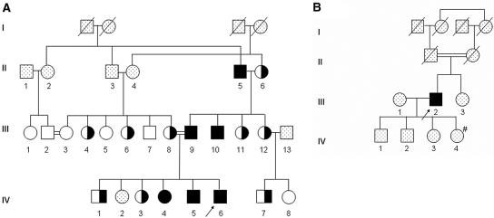
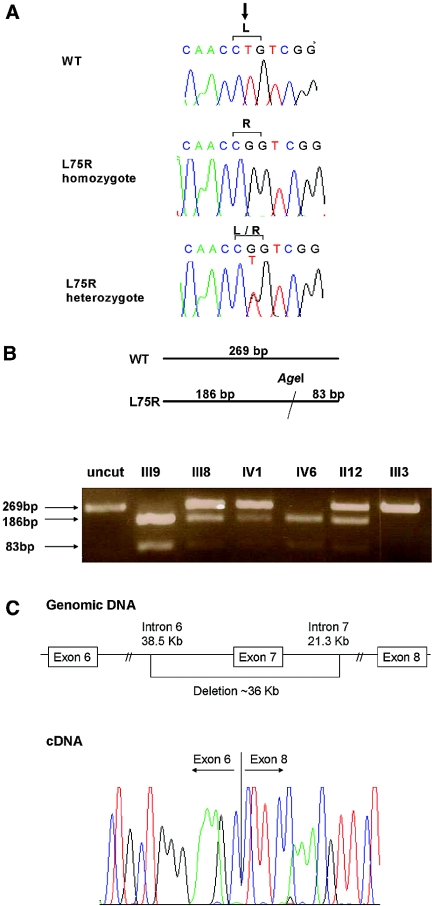
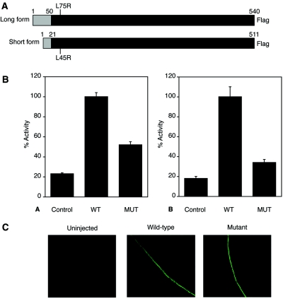
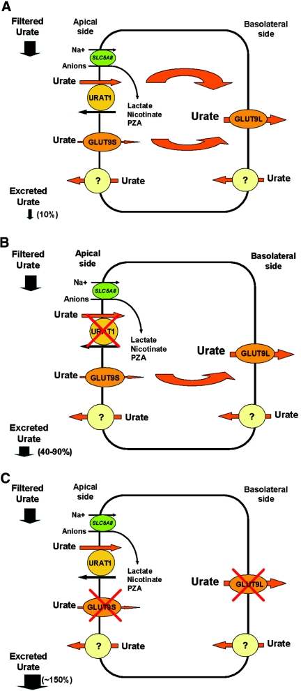
Hereditary hypouricemia may result from mutations in the renal tubular uric acid transporter URAT1. Whether mutation of other uric acid transporters produces a similar phenotype is unknown. We studied two families who had severe hereditary hypouricemia and did not have a URAT1 defect. We performed a genome-wide homozygosity screen and linkage analysis and identified the candidate gene SLC2A9, which encodes the glucose transporter 9 (GLUT9). Both families had homozygous SLC2A9 mutations: A missense mutation (L75R) in six affected members of one family and a 36-kb deletion, resulting in a truncated protein, in the other. In vitro, the L75R mutation dramatically impaired transport of uric acid. The mean concentration of serum uric acid of seven homozygous individuals was 0.17 +/- 0.2 mg/dl, and all had a fractional excretion of uric acid >150%. Three individuals had nephrolithiasis, and three had a history of exercise-induced acute renal failure. In conclusion, homozygous loss-of-function mutations of GLUT9 cause a total defect of uric acid absorption, leading to severe renal hypouricemia complicated by nephrolithiasis and exercise-induced acute renal failure. In addition to clarifying renal handling of uric acid, our findings may provide a better understanding of the pathophysiology of acute renal failure, nephrolithiasis, hyperuricemia, and gout.


Figures

Figure 1.
Figure 1.
Pedigrees of two unrelated consanguineous families with severe renal hypouricemia and SLC2A9 mutations. (A) Pedigree of family 1. (B) Pedigree of family 2. Solid symbols denote affected family members, open symbols denote unaffected family members, half-solid denote heterozygous family members, and dotted symbols denote family members who were not available for examination. Circles represent female family members, squares represent male family members, and crosses represent dead family members. Arrows indicate index patients. #Low serum UA (1.5 mg/dl), not available for genetic evaluation.
Figure 2.
Figure 2.
SLC2A9 mutations, found in two families with severe hereditary hypouricemia. (A) Missense mutation (c.T224G, p.L75R) found in the index patient of family 1 as compared with the sequences in a heterozygous family member and a healthy control subject (WT). (B) Restriction enzyme analysis of family 1, showing individuals with WT, homozygote, and heterozygote SLC2A9. The L75R mutation creates an AgeI recognition site that does not exist in the WT sequence. Digestion fragments of 269 and 83 bp are seen in mutated alleles. (C) Schematic presentation of the 36-kb deletion identified in the index patient of family 2 (top) and cDNA sequence of the transition from exon 6 to exon 8 (bottom).
Figure 3.
Figure 3.
(A) Schematic representation of the variants GLUT9L and GLUT9S showing the location of the mutation (L75R, L45R) found in family 1. (B) Reduced [8-14C]UA transport activity in oocytes injected with L75R mutant (MUT; A) or L46R mutant (MUT; B) compared with WT SLC2A9 mRNA. Oocytes were injected with control, WT, or mutant mRNA, and transport assays were performed at room temperature for 1 h, 2 d after injection. Activity is expressed as counts per minute of [8-14C]UA uptake, as a percentage of WT. The average of either three or four experiments is shown, and error bars represent SEs. (C) The L46R mutation does not significantly impair transport of GLUT9S to the plasma membrane. Immunodetection with an anti-GLUT9S antibody shows that the WT and L46R proteins are expressed at the plasma membrane, whereas fluorescence levels were undetectable in control oocytes or in the absence of GLUT9S antibody (data not shown).
Figure 4.
Figure 4.
A simplified model of UA handling by the proximal renal tubular cell, modified after Anzai et al. and Matsuo et al. (A) Normal physiology. (B) Loss-of-function of URAT1. (C) Loss-of-function of GLUT9 as a result of homozygous mutations.

References

    1. Wu XW, Lee CC, Muzny DM, Caskey CT: Urate oxidase: Primary structure and evolutionary implications. Proc Natl Acad Sci U S A 86: 9412–9416, 1989 - PMC - PubMed
    1. Feig DI, Kang DH, Johnson RJ: Uric acid and cardiovascular risk. N Engl J Med 359: 1811–1821, 2008 - PMC - PubMed
    1. Sperling O: Hereditary renal hypouricemia. Mol Genet Metab 89: 14–18, 2006 - PubMed
    1. Enomoto A, Kimura H, Chairoungdua A, Shigeta Y, Jutabha P, Cha SH, Hosoyamada M, Takeda M, Sekine T, Igarashi T, Matsuo H, Kikuchi Y, Oda T, Ichida K, Hosoya T, Shimokata K, Niwa T, Kanai Y, Endou H: Molecular identification of a renal urate anion exchanger that regulates blood urate levels. Nature 417: 447–452, 2002 - PubMed
    1. Ichida K, Hosoyamada M, Hisatome I, Enomoto A, Hikita M, Endou H, Hosoya T: Clinical and molecular analysis of patients with renal hypouricemia in Japan: Influence of URAT1 gene on urinary urate excretion. J Am Soc Nephrol 15: 164–173, 2004 - PubMed

Publication types

MeSH terms

Substances