. 2009 Oct;36(10):4425-32.
doi: 10.1118/1.3215926.

Evaluation of scatter effects on image quality for breast tomosynthesis

Affiliations

Evaluation of scatter effects on image quality for breast tomosynthesis

Gang Wu et al. Med Phys. 2009 Oct.

Abstract

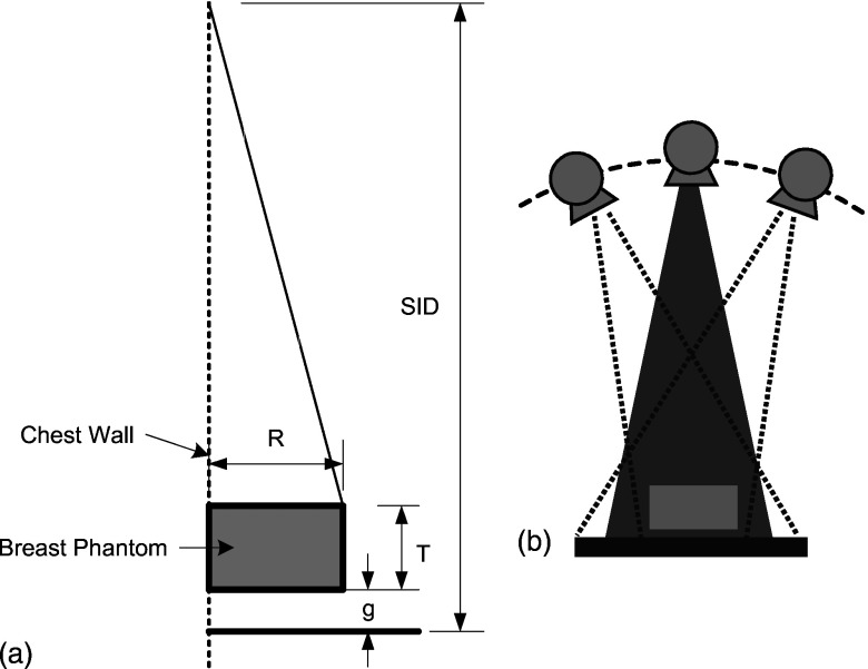
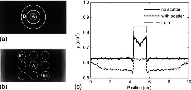
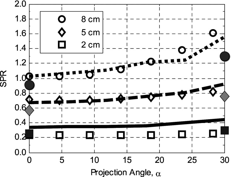
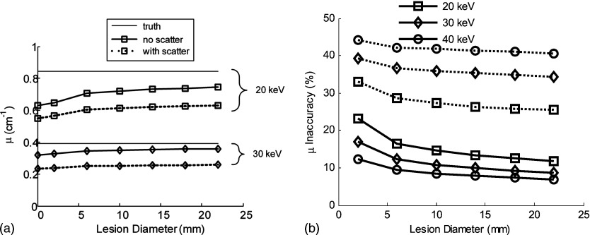
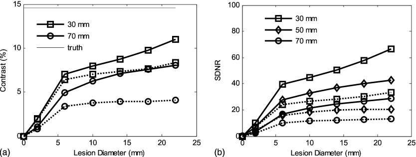
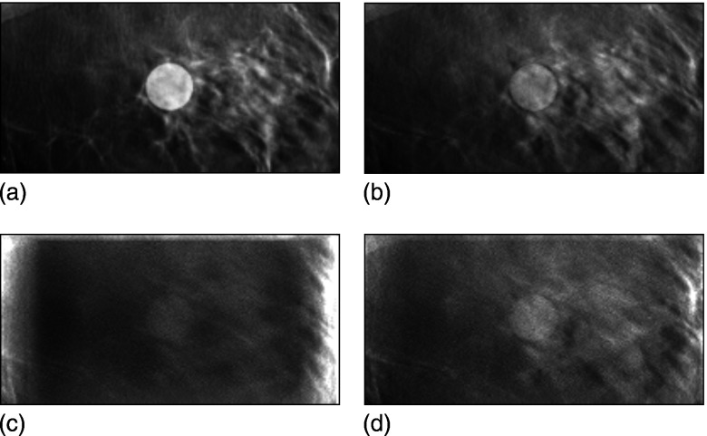
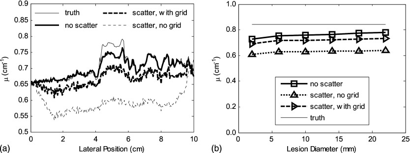
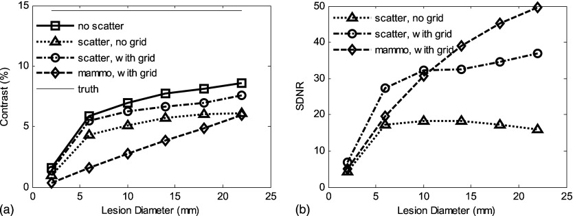
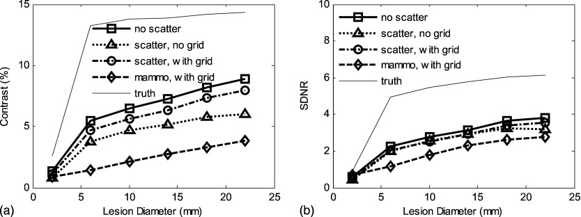
Digital breast tomosynthesis uses a limited number (typically 10-20) of low-dose x-ray projections to produce a pseudo-three-dimensional volume tomographic reconstruction of the breast. The purpose of this investigation was to characterize and evaluate the effect of scattered radiation on the image quality for breast tomosynthesis. In a simulation, scatter point spread functions generated by a Monte Carlo simulation method were convolved over the breast projection to estimate the distribution of scatter for each angle of tomosynthesis projection. The results demonstrate that in the absence of scatter reduction techniques, images will be affected by cupping artifacts, and there will be reduced accuracy of attenuation values inferred from the reconstructed images. The effect of x-ray scatter on the contrast, noise, and lesion signal-difference-to-noise ratio (SDNR) in tomosynthesis reconstruction was measured as a function of the tumor size. When a with-scatter reconstruction was compared to one without scatter for a 5 cm compressed breast, the following results were observed. The contrast in the reconstructed central slice image of a tumorlike mass (14 mm in diameter) was reduced by 30%, the voxel value (inferred attenuation coefficient) was reduced by 28%, and the SDNR fell by 60%. The authors have quantified the degree to which scatter degrades the image quality over a wide range of parameters relevant to breast tomosynthesis, including x-ray beam energy, breast thickness, breast diameter, and breast composition. They also demonstrate, though, that even without a scatter rejection device, the contrast and SDNR in the reconstructed tomosynthesis slice are higher than those of conventional mammographic projection images acquired with a grid at an equivalent total exposure.


Figures

Figure 1
Figure 1
(a) Imaging geometry: R—breast radius, T—breast thickness, and g—gap between the bottom of the phantom and the top of the detector. (b) Image acquisition positions for tomosynthesis: Partial isocentric geometry, in which the detector remains stationary and the x-ray tube moves around some center of rotation.
Figure 2
Figure 2
(a) Transaxial central slice of a tomosynthesis reconstruction for a breast phantom I, with a tumor at the center. (b) Transaxial central slice of a tomosynthesis reconstruction for a breast phantom I, without tumor. (c) Horizontal profile through the center of the phantom shows the reduction in the voxel value for both normal tissue and tumor.
Figure 3
Figure 3
Comparison of the SPR calculated in this simulation (data points: =8 cm breast thickness, =5 cm, =2 cm) with those previously reported by Sechopoulos et al. (Ref. 21) (dotted, dashed, and solid lines for the 8, 5, and 2 cm, respectively). Shaded symbols represent the corresponding results of Sechopoulos et al. without the compression plate in place.
Figure 4
Figure 4
(a) The voxel values (inferred linear attenuation coefficient compared to the true linear attenuation) of a tumor in the phantom, scatter-free reconstruction (solid lines), and with scatter (dashed lines) are illustrated for different energies as a function of lesion diameter. (b) Inaccuracy of μ (defined in text) vs lesion diameter in a 5-cm-thick phantom. The true values of μ are 0.629, 0.321, and 0.244cm1 at 20, 30, and 40 keV, respectively.
Figure 5
Figure 5
(a) The degrading effect of scatter radiation on contrast in tomosynthesis (phantom I) is illustrated for different breast thicknesses. (b) Scatter radiation markedly reduces the SDNR. Solid lines correspond to a scatter-free simulation and the dotted lines () correspond to simulations with scatter. Energy is 20 keV with constant entrance air kerma to the breast.
Figure 6
Figure 6
(a) Central slice of the original (truth) phantom II that has a tumor with a diameter of 14 mm at the center. Note that this image is from the original model data, not a reconstruction. (b) Corresponding tomosynthesis reconstructed slice, scatter free. (c) Reduced contrast and more noise were shown in the central slice of the tomosynthesis reconstruction with scattered radiation. (d) The use of a scatter reduction grid improves contrast and the accuracy of the voxel value. The windowing was set to be the same for all four images, with the level adjusted to yield approximately the same gray level in the background for each image.
Figure 7
Figure 7
(a) The cupping effect and reduction in accuracy due to scatter are illustrated in the profiles through the center of the tumor for phantom II. The potential value of a grid is also illustrated. (b) The degrading effect of scatter radiation on reconstructed voxel values is illustrated as a function of the lesion diameter.
Figure 8
Figure 8
For phantom I with uniform 50% fibroglandular tissue, 20 keV and 5 cm breast thickness: (a) Scatter-contaminated tomosynthesis slice has better contrast than mammographic CC view for a wide range of lesion diameters (from 2 to 22 mm). (b) Tomosynthesis slice has less SDNR than projection CC view; the use of grid improves the recovery of SDNR. Total incident air kerma was set to be 9.6 mJ/kg for all cases (yielding a mean glandular dose of approximately 3.1 mGy).
Figure 9
Figure 9
For phantom II (anatomic structures in background): (a) The scatter radiation reduces contrast in the tomosynthesis slice to a level comparable with a mammographic CC view. (b) Although degraded by scatter radiation, the SDNR in the tomosynthesis slice is higher than for the CC projection mammogram. The variation in contrast and SDNR for the “truth” data are caused by the fact that ROIs vary with the lesion diameter and the presence of a heterogeneous background. Incident air kerma was set the same as for the results shown in Fig. 8.

References

    1. Fahrig R., Mainprize J. G., Robert N., Rogers A., and Yaffe M. J., “Performance of glass fiber antiscatter devices at mammographic energies,” Med. Phys. 21(8), 1277–1282 (1994). 10.1118/1.597236 - DOI - PubMed
    1. Barnes J. E., “Characteristics and control of contrast in CT,” Radiographics 12(4), 825–837 (1992). - PubMed
    1. Shen S. Z., Bloomquist A. K., Mawdsley G. E., Yaffe M. J., and Elbakri I., “Effect of scatter and an antiscatter grid on the performance of a slot-scanning digital mammography system,” Med. Phys. 33(4), 1108–1115 (2006). 10.1118/1.2184445 - DOI - PubMed
    1. Boone J. M., Seibert J. A., Tang C. M., and Lane S. M., “Grid and slot scan scatter reduction in mammography: Comparison by using Monte Carlo techniques,” Radiology 222(2), 519–527 (2002). 10.1148/radiol.2222010491 - DOI - PubMed
    1. Barnes G. T. and Brezovich I. A., “The intensity of scattered radiation in mammography,” Radiology 126(1), 243–247 (1978). - PubMed

Publication types