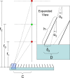
Nondestructive measurement of the grid ratio using a single image
- PMID: 19928103
- PMCID: PMC4034420
- DOI: 10.1118/1.3191245
Nondestructive measurement of the grid ratio using a single image
Abstract
The antiscatter grid is an essential part of modern radiographic systems. Since the introduction of the antiscatter grid, however, there have been few methods proposed for acceptance testing and verification of manufacturer-supplied grid specifications. The grid ratio (r) is an important parameter describing the antiscatter grid because it affects many other grid quality metrics, such as the contrast improvement ratio (K), primary transmission (Tp), and scatter transmission (Ts). Also, the grid ratio in large part determines the primary clinical use of the grid. To this end, the authors present a technique for the nondestructive measurement of the grid ratio of antiscatter grids. They derived an equation that can be used to calculate the grid ratio from a single off-focus flat field image by exploiting the relationship between grid cutoff and off-focus distance. The calculation can be performed by hand or with included analysis software. They calculated the grid ratios of several different grids throughout the institution, and afterward they destructively measured the grid ratio of a nominal r8 grid previously evaluated with the method. They also studied the sensitivity of the method to technical factors and choice of parameters. With one exception, the results for the grids found in the institution were in agreement with the manufacturer's specifications and international standards. The nondestructive evaluation of the r8 grid indicated a ratio of 7.3, while the destructive measurement indicated a ratio of 7.53 +/- 0.28. Repeated evaluations of the same grid yielded consistent results. The technique provides the medical physicist with a new tool for quantitative evaluation of the grid ratio, an important grid performance criterion. The method is robust and repeatable when appropriate choices of technical factors and other parameters are made.
Figures


Similar articles
-
Experimental evaluation of fiber-interspaced antiscatter grids for large patient imaging with digital x-ray systems.Phys Med Biol. 2007 Aug 21;52(16):4863-80. doi: 10.1088/0031-9155/52/16/010. Epub 2007 Jul 30. Phys Med Biol. 2007. PMID: 17671340
-
Efficiency of antiscatter grids for flat-detector CT.Phys Med Biol. 2007 Oct 21;52(20):6275-93. doi: 10.1088/0031-9155/52/20/013. Epub 2007 Oct 2. Phys Med Biol. 2007. PMID: 17921585
-
Improved image quality of cone beam CT scans for radiotherapy image guidance using fiber-interspaced antiscatter grid.Med Phys. 2014 Jun;41(6):061910. doi: 10.1118/1.4875978. Med Phys. 2014. PMID: 24877821
-
Effect of scatter and an antiscatter grid on the performance of a slot-scanning digital mammography system.Med Phys. 2006 Apr;33(4):1108-15. doi: 10.1118/1.2184445. Med Phys. 2006. PMID: 16696488
-
Contrast and scatter in x-ray imaging.Radiographics. 1991 Mar;11(2):307-23. doi: 10.1148/radiographics.11.2.2028065. Radiographics. 1991. PMID: 2028065 Review.
References
-
- Bucky G., “Über die ausschaltung der im objekt entstehenden sekundärstrahlen bei röntgenanfnahmen,” Verh. Dtsch. Ront. Ges. 9, 30 (1913).
-
- “Specification, acceptance testing and quality control of diagnostic x-ray imaging equipment,” 1991 AAPM Summer School Proceedings, Seibert J. A., Barnes G. T., and Gould R. (American Institute of Physics, Santa Cruz, CA, 1991).
-
- International Electrotechnical Commission, Diagnostic X-Ray Imaging Equipment—Characteristics of General Purpose and Mammographic Anti-scatter Grids (IEC, Geneva, 2001).
-
- “Quality assurance for diagnostic imaging,” National Council on Radiation Protection and Measurements Report No 99 (NCRP, Bethesda, MD, 1988).
-
- “Quality control in diagnostic radiology,” American Association of Physicists in Medicine Report No. 74 (Medical Physics, Madison, WI, 2002).
Publication types
MeSH terms
LinkOut - more resources
Full Text Sources

