

Repeatability of layered corneal pachymetry with the artemis very high-frequency digital ultrasound arc-scanner
- PMID: 19928698
- PMCID: PMC4464782
- DOI: 10.3928/1081597X-20091105-01
Repeatability of layered corneal pachymetry with the artemis very high-frequency digital ultrasound arc-scanner
Abstract
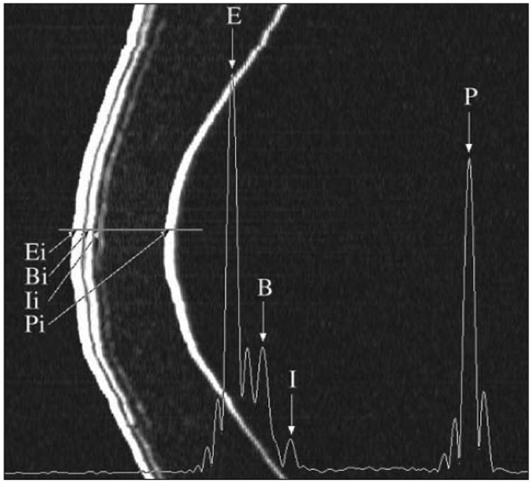
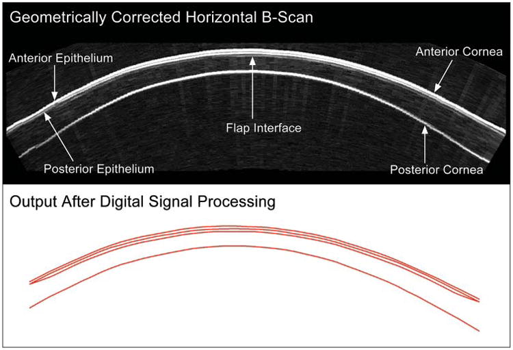
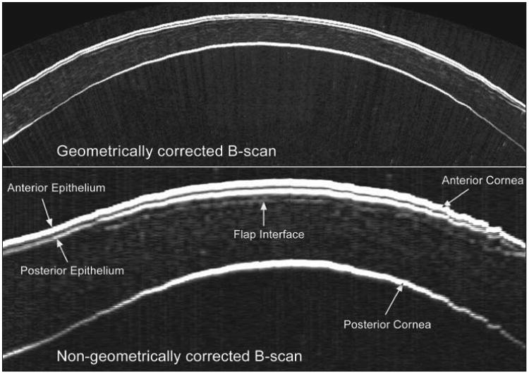
Purpose: To assess the three-dimensional repeatability of thickness measurements for epithelium, stroma, cornea, flap, and residual stromal bed using the Artemis very high-frequency (VHF) digital ultrasound arc-scanner (ArcScan Inc).
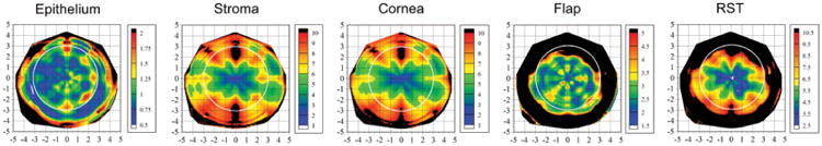
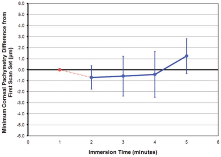
Methods: Five consecutive measurements were obtained for 10 eyes of 10 patients 1 year after LASIK using the Artemis VHF digital ultrasound arc-scanner across the central 10-mm diameter of the cornea. Repeatability analysis was performed for thickness measurements for each corneal layer-epithelium, stroma, cornea, flap, and residual stromal bed. The standard deviation of repeated measurements (point-repeatability) was calculated for each measurement location in 0.1-mm steps for the 10×10-mm matrix. The pooled standard deviation of the point-repeatability for each measurement location within the central 1-, 2-, and 3-mm radius was calculated (region-repeatability). The corneal thickness of the baseline scan set was compared to that of subsequent scan sets within the same session and plotted over time to assess any possible hydration effects of the immersion technique.
Results: The repeatability at the corneal vertex was 0.58 μm for epithelium, 1.78 μm for stroma, 1.68 μm for cornea, 1.68 μm for flap, and 2.27 μm for residual stromal bed. The region-repeatability within the central 1-mm radius was 1.01 μm for epithelium, 3.44 μm for stroma, 3.35 μm for cornea, 2.81 μm for flap, and 3.97 μm for residual stromal bed. The mean difference in corneal thickness from the baseline value was within 1.25 μm for each of the subsequent four scan sets over a 5-minute immersion period.
Conclusions: Layered pachymetry of the epithelium, stroma, cornea, flap, and residual stromal bed showed high repeatability with the Artemis VHF digital ultrasound arc-scanner. The high repeatability validates the use of the Artemis for in vivo layered pachymetry.
Copyright 2010, SLACK Incorporated.
Figures
References
-
- Reinstein DZ, Silverman RH, Coleman DJ. High-frequency ultrasound measurement of the thickness of the corneal epithelium. Refract Corneal Surg. 1993;9(5):385–387. - PubMed
-
- Reinstein DZ, Silverman RH, Trokel SL, Coleman DJ. Corneal pachymetric topography. Ophthalmology. 1994;101(3):432–438. - PubMed
-
- Reinstein DZ, Silverman RH, Rondeau MJ, Coleman DJ. Epithelial and corneal thickness measurements by high-frequency ultrasound digital signal processing. Ophthalmology. 1994;101(1):140–146. - PubMed
-
- Reinstein DZ, Silverman RH, Sutton HF, Coleman DJ. Very high-frequency ultrasound corneal analysis identifies anatomic correlates of optical complications of lamellar refractive surgery: anatomic diagnosis in lamellar surgery. Ophthalmology. 1999;106(3):474–482. - PubMed
-
- Reinstein DZ, Patel S, Aslanides IM, Silverman RH, Coleman DJ. Epithelial lenticular types of human cornea: classification and analysis of influence on PRK. Ophthalmology. 1995;102(Suppl):156.
Publication types
MeSH terms
Grants and funding
LinkOut - more resources
Full Text Sources
Other Literature Sources
Medical
