. 2010 Jan 14;53(1):345-56.
doi: 10.1021/jm901325e.

Design of O-acetylserine sulfhydrylase inhibitors by mimicking nature

Affiliations

Design of O-acetylserine sulfhydrylase inhibitors by mimicking nature

Enea Salsi et al. J Med Chem. .

Abstract

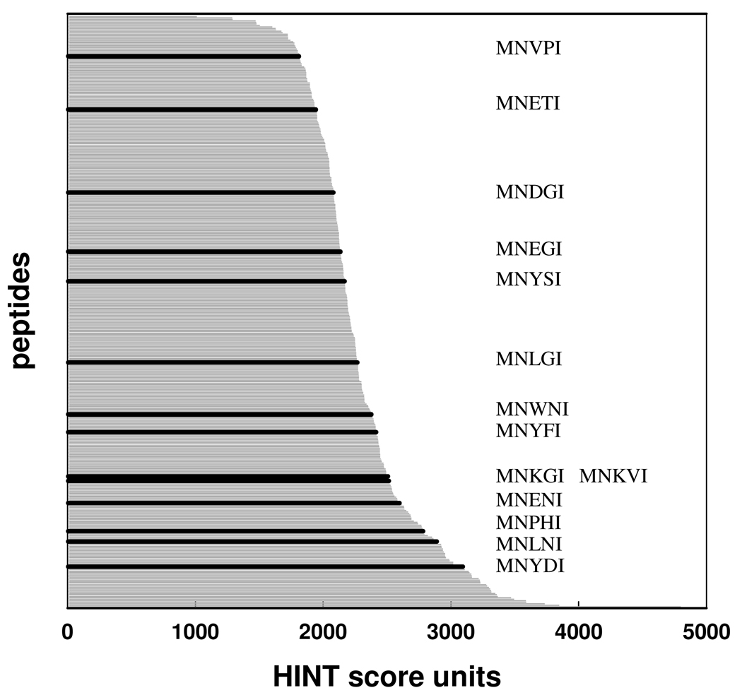
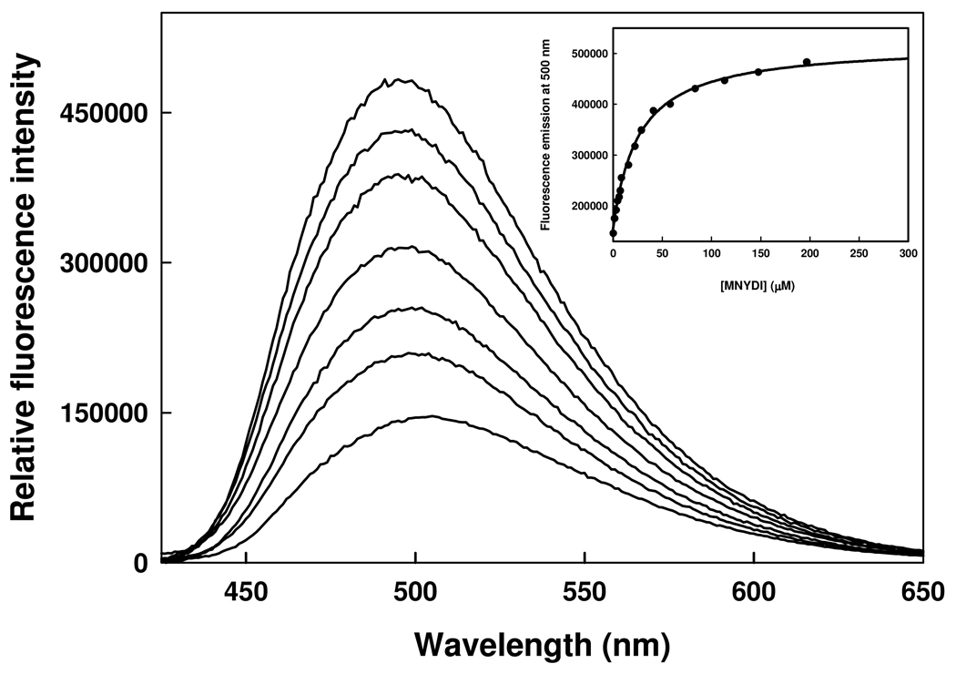
The inhibition of cysteine biosynthesis in prokaryotes and protozoa has been proposed to be relevant for the development of antibiotics. Haemophilus influenzae O-acetylserine sulfhydrylase (OASS), catalyzing l-cysteine formation, is inhibited by the insertion of the C-terminal pentapeptide (MNLNI) of serine acetyltransferase into the active site. Four-hundred MNXXI pentapeptides were generated in silico, docked into OASS active site using GOLD, and scored with HINT. The terminal P5 Ile accounts for about 50% of the binding energy. Glu or Asp at position P4 and, to a lesser extent, at position P3 also significantly contribute to the binding interaction. The predicted affinity of 14 selected pentapeptides correlated well with the experimentally determined dissociation constants. The X-ray structure of three high affinity pentapeptide-OASS complexes were compared with the docked poses. These results, combined with a GRID analysis of the active site, allowed us to define a pharmacophoric scaffold for the design of peptidomimetic inhibitors.


Figures

Figure 1
Figure 1
a. Ribbon-tube representation of HiSAT (PDB code 1sst). The C-terminal region (aa 241–267), including the peptide complexed with HiOASS-A (aa 258–267) is not shown in the picture since this region was not crystallographically detected. b. Ribbon-tube representation of HiOASS-A complexed with the SAT C-terminal tetrapeptide (PDB code 1y7l). c. HiOASS-A binding pocket. The SAT C-terminal tetrapeptide is represented in yellow sticks. The HiOASS-A residues forming H-bonds (white dashed lines) with the SAT peptide are shown in transparent white sticks. The binding pocket surface is displayed as a function of the lipophilic potential. Lipophilic regions are colored brown, whereas the polar cleft regions are progressively colored green, cyan and blue. Only essential hydrogens are shown.
Figure 2
Figure 2
HINT score fraction (expressed as a percentage of the total HINT score value) assigned to the interaction between the different SAT C-terminal residues and the OASS-A binding pocket in the crystallographic HiOASS-A decapeptide complex.
Figure 3
Figure 3
HINT score values assigned to the interaction of the 400 MNXXI docked peptides and the HiOASS-A binding pocket. The HINT scores (grey lines) are reported in decreasing order. The pentapeptides experimentally tested are shown in black
Figure 4
Figure 4
Binding of MNYDI to HiOASS. Fluorescence emission spectra upon excitation at 412 nm (slitex= 4 nm, slitem= 4 nm) of a solution containing 1 µM HiOASS and increasing concentrations of MNYDI in 100 mM Hepes buffer, pH 7, at 20°C. Inset. Dependence of the fluorescence emission intensity at 500 nm on the peptide concentration. Line through data points is the fit to a binding isotherm with Kd of 25.8 ± 1.7 µM.
Figure 5
Figure 5
Correlation between the experimental pKd and the computational HINT scores for the 14 selected pentapeptides. The correlation is characterized by the equation pKdiss = 0.0018 Htotal – 0.60 with an r2 of 0.65 and a standard error of 0.54 pKdiss units.
Figure 6
Figure 6
X-ray structures of MNWNI, MNYDI and MNENI bound to HiOASS and comparison with the conformations generated from docking/scoring. a. Superposition of the crystallographically determined structures of MNLNI (yellow), MNWNI (green), MNYDI (light blue) and MNENI (orange) within the HiOASS active site. b. MNWNI conformations generated from docking/scoring and determined crystallographically are displayed in magenta and green, respectively. c. MNYDI conformations generated from docking/scoring and determined crystallographically are displayed in magenta and light blue, respectively. d. MNENI conformations generated from docking/scoring and determined crystallographically are displayed in magenta and orange, respectively. For b–d the HiOASS active site is represented by a Connolly surface built with Sybyl MOLCAD tools and colored as a depth function. External protruding regions are colored blue, while cavities and clefts are progressively colored green, yellow and orange.
Figure 7
Figure 7
Mapping of the energetic contribution of the 400 pentapeptides. The black line represents the HINT scores attributed to the residues at position P3, the red line represents the sum of HINT scores at positions P3 and P4, and the cyan line represents the sum of HINT scores at positions P3, P4 and P5. The dotted line is the zero value.
Figure 8
Figure 8
GRID Molecular Interaction Fields calculated for the HiOASS active site. The red, blue and green-colored contours correspond to the Molecular Interaction Fields calculated for H-bond acceptor (O), H-bond donor (N1) and hydrophobic (DRY) probes. The active site surface is represented as a function of the cavity depth while the wild type crystallographic SAT C-terminal tetrapeptide is shown in transparent capped sticks.
Scheme 1
Scheme 1
Assimilatory pathway of sulfur incorporation into cysteine in enteric bacteria. Enzymes are shown in bold italics. Inhibition (formula image) and activation (formula image) of enzyme activities are shown in colour. Cysteine inhibits SAT and SAT inhibits OASS-A. OASS-A activates ATP sulfurylase.
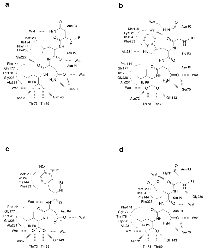
Scheme 2
Scheme 2
Interactions between the HiOASS active site residues and the peptides as derived from the crystallographic structure for complexes with: a. MNLNI; b. MNWNI; c. MNYDI; d. MNENI.

References

    1. de Lencastre H, Oliveira D, Tomasz A. Antibiotic resistant Staphylococcus aureus: a paradigm of adaptive power. Curr. Opin. Microbiol. 2007;10:428–435. - PMC - PubMed
    1. Livermore DM. The need for new antibiotics. Clin. Microbiol. Infect. 2004;10 Suppl 4:1–9. - PubMed
    1. Nikaido H. Multidrug resistance in bacteria. Annu. Rev. Biochem. 2009;78:119–146. - PMC - PubMed
    1. Conly J, Johnston B. Where are all the new antibiotics? The new antibiotic paradox. Can. J. Infect. Dis. Med. Microbiol. 2005;16:159–160. - PMC - PubMed
    1. Spellberg B, Powers JH, Brass EP, Miller LG, Edwards JE., Jr. Trends in antimicrobial drug development: implications for the future. Clin. Infect. Dis. 2004;38:1279–1286. - PubMed

Publication types

LinkOut - more resources