. 2009 Dec 29;48(51):12191-201.
doi: 10.1021/bi901640e.

Structure, stability, and interaction of the fibrin(ogen) alphaC-domains

Affiliations

Structure, stability, and interaction of the fibrin(ogen) alphaC-domains

Galina Tsurupa et al. Biochemistry. .

Abstract

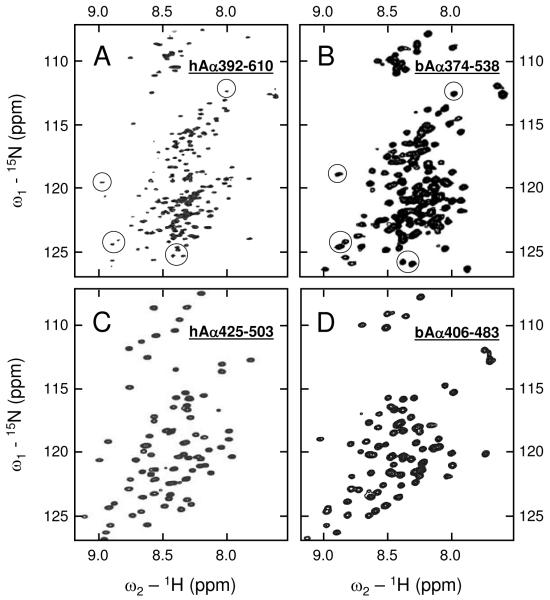
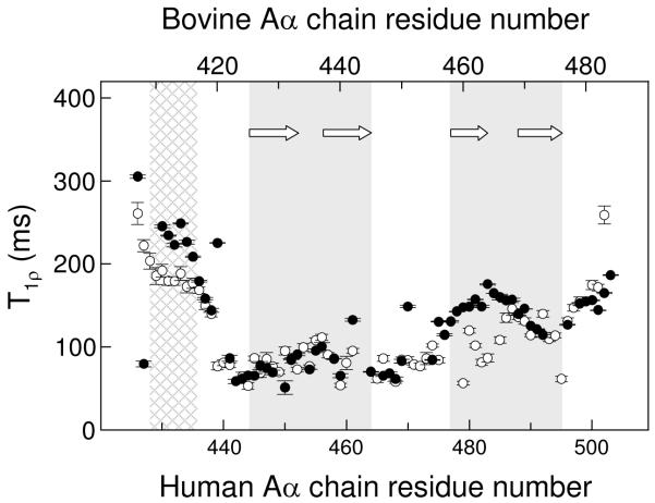
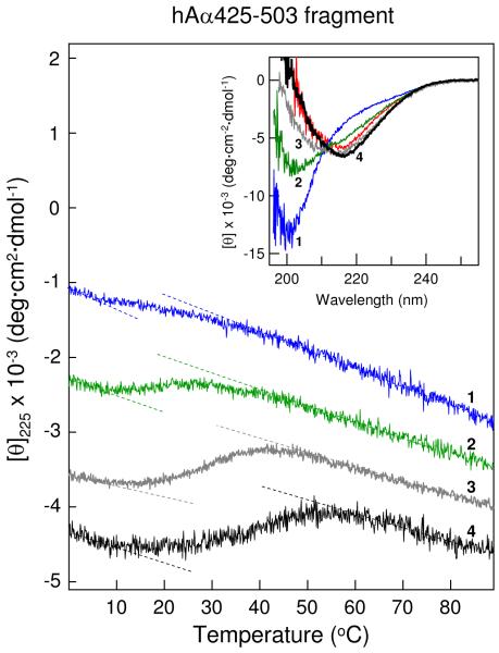
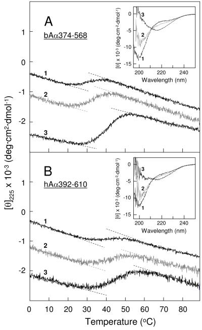
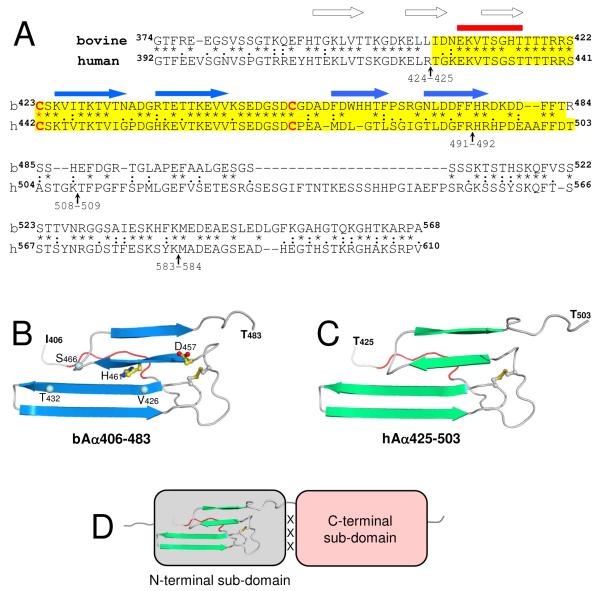
Our recent study established the NMR structure of the recombinant bAalpha406-483 fragment corresponding to the NH(2)-terminal half of the bovine fibrinogen alphaC-domain and revealed that at increasing concentrations this fragment forms oligomers (self-associates). The major goals of the study presented here were to determine the structure and self-association of the full-length human fibrinogen alphaC-domains. To accomplish these goals, we prepared a recombinant human fragment, hAalpha425-503, homologous to bovine bAalpha406-483, and demonstrated using NMR, CD, and size-exclusion chromatography that its overall fold and ability to form oligomers are similar to those of bAalpha406-483. We also prepared recombinant hAalpha392-610 and bAalpha374-568 fragments corresponding to the full-length human and bovine alphaC-domains, respectively, and tested their structure, stability, and ability to self-associate. Size-exclusion chromatography revealed that both fragments form reversible oligomers in a concentration-dependent manner. Their oligomerization was confirmed in sedimentation equilibrium experiments, which also established the self-association affinities of these fragments and revealed that the addition of each monomer to assembling alphaC-oligomers substantially increases the stabilizing free energy. In agreement, unfolding experiments monitored by CD established that self-association of both fragments results in a significant increase in their thermal stability. Analysis of CD spectra of both fragments revealed that alphaC self-association results in an increase in the level of regular structure, implying that the COOH-terminal half of the alphaC-domain adopts an ordered conformation in alphaC-oligomers and that this domain contains two independently folded subdomains. Altogether, these data further clarify the structure of the human and bovine alphaC-domains and the molecular mechanism of their self-association into alphaC-polymers in fibrin.


Figures

Figure 1
Figure 1
1H-15N HSQC NMR spectra of the hAα392-610 (panel A), bAα374-538 (panel B), hAα425-503 (panel C), and bAα406-483 (panel D) fragments. Spectrum A was taken with slightly smaller spectral width in the 15N dimension in an attempt to enhance peak resolution. Several peaks assigned to the first disulfide-linked β-hairpin in bAα374-538 are circled in panel B; those occurring at similar positions in hAα392-610 are circled in panel A.
Figure 2
Figure 2
15N backbone relaxation data demonstrating similarity in relaxation and dynamics of the human hAα425-503 and bovine bAα406-483 fragments. T1p relaxation data for hAα425-503 and bAα406-483 are shown by filled and empty circles, respectively; vertical bars represent experimental errors. The regions corresponding to the previously identified first and second β-hairpins in the bovine bAα406-483 fragment (19) are shaded in gray, β-strands forming these β-hairpins are shown by arrows; the NH2-terminal region of slower motion in this fragment (19) is shown by cross-hatched pattern.
Figure 3
Figure 3
CD-detected thermal unfolding of the hAα425-503 fragment. The unfolding experiments were performed in 20 mM Tris buffer, pH 7.4, containing 0.15 M NaCl at 3.2 mg/mL hAα425-503 (blue curve 1) and in the same buffer containing either 1 M NaCl at 3.2 mg/mL hAα425-503 (green curve 2) or 2 M NaCl at 1.6 mg/mL and 3.4 mg/mL hAα425-503 (gray curve 3 and black curve 4, respectively). The unfolding curves have been arbitrary shifted along the vertical axis to improve visibility; the dashed straight lines represent linear extrapolations of the CD values before and after transitions to highlight their sigmoidal character. Inset shows CD spectra of the hAα425-503 fragment obtained in the above mentioned conditions; the numbering and color coding of these spectra correspond to those of the unfolding curves. The CD spectrum of the bovine bAα406-483 fragment at 3.0 mg/ml in Tris-buffer, pH 7.4, containing 0.15 M NaCl is shown in red for comparison. All spectra were obtained at 4 °C.
Figure 4
Figure 4
Size-exclusion chromatography of the hAα425-503 fragment performed in various conditions. Panels A and B show elution profiles of hAα425-503 in Tris buffer, pH 7.4, containing 0.15 M NaCl, at 3.2 and 6.3 mg/mL, respectively. Panels C and D show elution profiles of hAα425-503 in the same buffer containing 2 M NaCl at 1.6 and 3.4 mg/mL, respectively. All experiments were performed at 4 °C using the Superdex 75 column; arrows indicate the free volume of the column.
Figure 5
Figure 5
CD-detected thermal unfolding of the full-length bovine and human αC-domain fragments. The unfolding of the bovine bAα374-568 fragment (panel A) was performed in 20 mM Tris-buffer, pH 7.4, containing 0.15 M NaCl, at the fragment concentrations of 1.8 and 3.7 mg/mL (curve 1 and 2, respectively), and in the same buffer containing 2 M NaCl at the fragment concentration of 3.8 mg/mL (curve 3). The unfolding of the human hAα392-610 fragment (panel B) was performed in Tris-buffer, pH 7.4, containing 0.15 M NaCl, at the fragment concentrations of 1.9 and 3.8 mg/mL (curve 1 and 2, respectively), and in the same buffer containing 2 M NaCl at the fragment concentration of 4.0 mg/mL (curve 3). The unfolding curves have been arbitrary shifted along the vertical axis to improve visibility; the dashed straight lines represent linear extrapolations of the CD values before and after transitions to highlight their sigmoidal character. Insets in both panels shows CD spectra of the corresponding fragments obtained in the above mentioned conditions at 4 °C; the numbering of these spectra correspond to that of the unfolding curves.
Figure 6
Figure 6
Analysis of oligomerization of the human hAα392-610 (panels A-C) and bovine bAα374-568 (panels D-F) fragments by analytical ultracentrifugation at 6,000 rpm (open circles) and 8,000 rpm (grey triangles). The experiments were performed in TBS at three concentrations of the fragments, 2.0, 4.1 and 8.1 mg/mL (panels A, B and C, respectively), and 1.8, 3.8 and 7.2 mg/mL (panels D, E and F, respectively). The data presented in panels A and D were obtained in a 12 mm path length cell, whereas those presented in panels B, C, E and F were obtained at higher concentrations in 3 mm path length cells. Sedimentation equilibrium data for both fragments obtained at all three concentrations demonstrate oligomerization. HeteroAnalysis of the data obtained for the hAα392-610 (A-C) and bAα374-568 (D-F) fragments yielded Mw = 266,910 and 179,029, respectively; the solid lines describe an isodesmic self-association model with Ka = 8.32 × 10⁴ M−1 and 2.61 × 10⁴ M−1 for hAα392-610 and bAα374-568, respectively. This model accounts for both rotor speed and concentration-dependence as evidenced by the correspondence between data and fitted lines in all panels.
Figure 7
Figure 7
Structural organization of the fibrinogen αC-domain. Panel A, alignment of the human and bovine fibrinogen αC-domain sequences performed as in (27). The degree of conservation is represented by asterisks, colons and dots, which denote identical residues, conserved and semi-conserved substitutions, respectively; the sequences corresponding to the bovine bAα406-483 and human hAα425-503 fragments are highlighted in yellow; vertical arrows indicate the identified plasmin cleavage sites (1); locations of the NH2-terminal region of slower motion (see Figure 2 and ref. 19) and β-sheet strands identified in bAα406-483 (19) are shown by red horizontal bar and blue horizontal arrows, respectively. Location of predicted β-strands in the bovine Aα374-422 region is shown by empty arrows. Panel B, the ribbon diagram of the bovine bAα406-483 fragment based upon its NMR structure (19); arrows indicate β-strands, the region of slower motion is shown in red. Asn457 and His461 missing in the human sequence are shown by balls and sticks; the location of Val426, Thr432, and Ser461 mentioned in the text are shown by blue balls. Panel C, the homology model of the human hAα425-503 fragment built based on the 3D structure of bovine bAα406-483. Panel D, schematic representation of the αC-domain consisting of the N-terminal sub-domain, which includes the previously identified β-sheet (19), and the C-terminal sub-domain whose structure is not established yet; interaction between these domains (see text) is denoted by ‘XXX’. Figures on panels B and C were prepared using PyMOL (38).

References

    1. Henschen A, McDonagh J. Fibrinogen, fibrin and factor XIII. In: Zwaal RFA, Hemker HC, editors. Blood Coagulation. Elsievier Science Publishers; Amsterdam: 1986. pp. 171–241.
    1. Medved LV, Gorkun OV, Privalov PL. Structural organization of C-terminal parts of fibrinogen Aα-chains. FEBS Lett. 1983;160:291–295. - PubMed
    1. Erickson HP, Fowler WE. Electron microscopy of fibrinogen, its plasmic fragments and small polymers. Ann. N. Y. Acad. Sci. 1983;408:146–163. - PubMed
    1. Weisel JW, Medved L. The structure and function of the αC domains of fibrinogen. Ann. N. Y. Acad. Sci. 2001;936:312–327. - PubMed
    1. Medved LV, Gorkun OV, Manyakov VF, Belitser VA. The role of fibrinogen αC-domains in the fibrin assembly process. FEBS Lett. 1985;181:109–112. - PubMed

Publication types

LinkOut - more resources