Review
. 2009 Dec 29;48(51):12062-80.
doi: 10.1021/bi901739t.

Ca(2+) channels on the move

Affiliations
Free PMC article
Review

Ca(2+) channels on the move

Colin W Taylor et al. Biochemistry. .
Free PMC article

Erratum in

  • Biochemistry. 2010 Jun 22;49(24):5082

Abstract

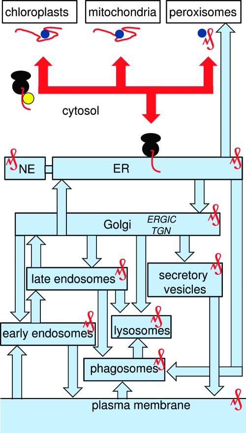
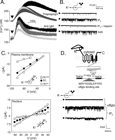
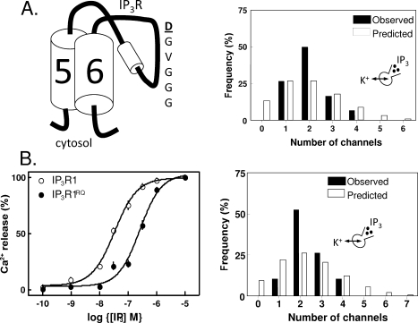
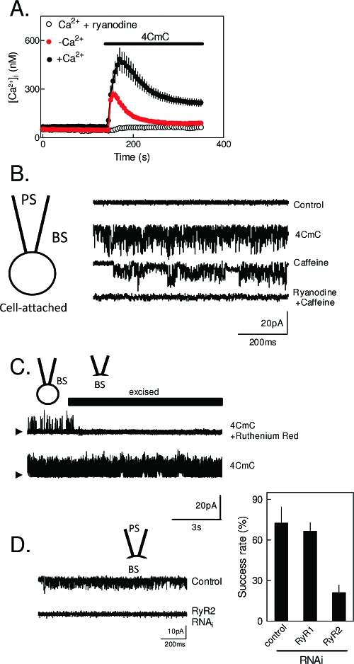
The versatility of Ca(2+) as an intracellular messenger derives largely from the spatial organization of cytosolic Ca(2+) signals, most of which are generated by regulated openings of Ca(2+)-permeable channels. Most Ca(2+) channels are expressed in the plasma membrane (PM). Others, including the almost ubiquitous inositol 1,4,5-trisphosphate receptors (IP(3)R) and their relatives, the ryanodine receptors (RyR), are predominantly expressed in membranes of the sarcoplasmic or endoplasmic reticulum (ER). Targeting of these channels to appropriate destinations underpins their ability to generate spatially organized Ca(2+) signals. All Ca(2+) channels begin life in the cytosol, and the vast majority are then functionally assembled in the ER, where they may either remain or be dispatched to other membranes. Here, by means of selective examples, we review two issues related to this trafficking of Ca(2+) channels via the ER. How do cells avoid wayward activity of Ca(2+) channels in transit as they pass from the ER via other membranes to their final destination? How and why do some cells express small numbers of the archetypal intracellular Ca(2+) channels, IP(3)R and RyR, in the PM?


Figures

Figure 1
Figure 1
Targeting channels to different membranes from the cytosol. Protein synthesis begins in the cytosol before targeting to specific organelles (red lines). This can occur post-translationally as unfolded proteins (mitochondria and chloroplasts) or fully folded proteins (peroxisomes), each protein protected by chaperones (blue circles). Most ER targeting occurs cotranslationally, mediated by SRP (yellow) binding to a signal sequence. Once inserted into the ER membrane, proteins assume the topology that they will retain through all subsequent trafficking steps. Some proteins may pass to the nuclear envelope (NE) or be exported directly from the ER to peroxisomes, but all other proteins pass via the ER−Golgi intermediate compartment (ERGIC) to the Golgi before being sorted to various destinations from the trans-Golgi network (TGN). Routes to and from the PM are shown.
Figure 2
Figure 2
Expression of IP3 receptors in the plasma membrane of DT40 cells. (A) Thapsigargin (top) stimulates Ca2+ release (palest line, in Ca2+-free medium) and Ca2+ entry (black line, in Ca2+-containing medium). The latter, SOCE, is entirely blocked by 300 nM GdCl3 (gray line). The bottom panel shows that activation of the BCR with anti-IgM stimulates both Ca2+ release and Ca2+ entry, but the latter is only partially inhibited by GdCl3. (B) Whole-cell patch-clamp recoding from DT40 cells with IP3, IP3 with heparin, or adenophostin A in the patch pipette. The holding potential was −100 mV; arrowheads denote the closed state. (C) Current−voltage (iV) relationships for the IP3-stimulated currents recorded from the PM or nuclear envelope of DT40-KO cells stably transfected with wild-type IP3R1 (R1) or IP3R1 with mutants in the putative pore (G2547A, R1GA; V2548I, R1VI). The point mutations similarly affected γK of the IP3-activated currents in both settings. (D) The six TMDs of a single IP3R subunit are shown to highlight the putative selectivity filter (sf) and the engineered αBgtx-binding site. In whole-cell patch-clamp recordings from DT40 cells expressing IP3R1 with this αBgtx-binding site, intracellular IP3 stimulated channel openings, and both Po and γK were increased by extracellular αBgtx. Reproduced from ref (65) with permission. Copyright 2006. American Academy for the Advancement of Science.
Figure 3
Figure 3
Counting IP3 receptors into the plasma membrane. (A) A point mutation within the putative pore region of IP3R1 (D2550A, highlighted) causes luminal/extracellular Ca2+ to block the channel, but it does not prevent cells from reliably counting IP3R into the PM. The histogram shows the observed and predicted (from the Poisson distribution) numbers of functional IP3R detected in each cell and establishes that IP3R are not randomly inserted into the PM. (B) A point mutation within the IP3-binding core (R568Q, IP3R1RQ) reduces the binding affinity of the IP3R for IP3 by 10-fold, evidenced by radioligand binding analyses (not shown) and the 10-fold decrease in the sensitivity of Ca2+ release to IP3 (left). The reduced sensitivity to IP3 does not impair the reliability with which IP3R are functionally expressed in the PM (right). Reproduced with permission from ref (140). Copyright 2008. American Society for Biochemistry and Molecular Biology.
Figure 4
Figure 4
Functional ryanodine receptors in the plasma membrane of RINm5F insulinoma cells. (A) Ca2+ signals evoked in populations of cells by 4CmC (1 mM) with or without prior treatment with ryanodine (400 μM) and in either normal or Ca2+-free medium. The results are consistent with activation of RyR causing both Ca2+ release and Ca2+ entry. (B) Cell-attached recordings from cells with cesium methanesulfonate in both bathing (BS) and pipette (PS) solutions at a holding potential of −100 mV. Caffeine (1 mM), 4CmC (1 mM), or ryanodine (400 μM) was included in BS as indicated. Arrowheads denote the closed state. (C) 4CmC activates channels in the cell-attached mode, which are then rapidly inhibited when the patch is excised into BS containing the membrane-impermeant inhibitor of RyR, ruthenium red (10 μM). (D) Selective inhibition of RyR2 expression using RNAi attenuates the electrical activity evoked by 4CmC. Typical records for control and RyR2-RNAi-treated cells are shown, and the success rate for detecting 4CmC-activated channels in the PM is shown for mock-transfected cells or cells transfected with RNAi for RyR1 or RyR2. Reproduced with permission from ref (185). Copyright 2009. American Society for Biochemistry and Molecular Biology.

References

    1. Berridge M. J.; Bootman M. D.; Roderick H. L. (2003) Calcium signalling: Dynamics, homeostasis and remodelling. Nat. Rev. Mol. Cell Biol. 4, 517–529. - PubMed
    1. Berridge M. J.; Lipp P.; Bootman M. D. (2000) The versatility and universality of calcium signalling. Nat. Rev. Mol. Cell Biol. 1, 11–21. - PubMed
    1. Xiong T. C.; Bourque S.; Lecourieux D.; Amelot N.; Grat S.; Briere C.; Mazars C.; Pugin A.; Ranjeva R. (2006) Calcium signaling in plant cell organelles delimited by a double membrane. Biochim. Biophys. Acta 1763, 1209–1215. - PubMed
    1. Kirichok Y.; Krapavinsky G.; Clapham D. E. (2004) The mitochondrial calcium uniporter is a highly selective ion channel. Nature 427, 360–364. - PubMed
    1. Toyoshima C. (2008) Structural aspects of ion pumping by Ca2+-ATPase of sarcoplasmic reticulum. Arch. Biochem. Biophys. 476, 3–11. - PubMed

Publication types

MeSH terms

Substances