. 2010 Jul;25(7):1680-94.
doi: 10.1359/jbmr.091116.

Origin matters: differences in embryonic tissue origin and Wnt signaling determine the osteogenic potential and healing capacity of frontal and parietal calvarial bones

Affiliations

Origin matters: differences in embryonic tissue origin and Wnt signaling determine the osteogenic potential and healing capacity of frontal and parietal calvarial bones

Natalina Quarto et al. J Bone Miner Res. 2010 Jul.

Abstract

Calvarial bones arise from two embryonic tissues, namely, the neural crest and the mesoderm. In this study we have addressed the important question of whether disparate embryonic tissue origins impart variable osteogenic potential and regenerative capacity to calvarial bones, as well as what the underlying molecular mechanism(s). Thus, by performing in vitro and in vivo studies, we have investigated whether differences exist between neural crest-derived frontal and paraxial mesodermal-derived parietal bone. Of interest, our data indicate that calvarial bone osteoblasts of neural crest origin have superior potential for osteogenic differentiation. Furthermore, neural crest-derived frontal bone displays a superior capacity to undergo osseous healing compared with calvarial bone of paraxial mesoderm origin. Our study identified both in vitro and in vivo enhanced endogenous canonical Wnt signaling in frontal bone compared with parietal bone. In addition, we demonstrate that constitutive activation of canonical Wnt signaling in paraxial mesodermal-derived parietal osteoblasts mimics the osteogenic potential of frontal osteoblasts, whereas knockdown of canonical Wnt signaling dramatically impairs the greater osteogenic potential of neural crest-derived frontal osteoblasts. Moreover, fibroblast growth factor 2 (FGF-2) treatment induces phosphorylation of GSK-3beta and increases the nuclear levels of beta-catenin in osteoblasts, suggesting that enhanced activation of Wnt signaling might be mediated by FGF. Taken together, our data provide compelling evidence that indeed embryonic tissue origin makes a difference and that active canonical Wnt signaling plays a major role in contributing to the superior intrinsic osteogenic potential and tissue regeneration observed in neural crest-derived frontal bone.


Figures

Fig. 1
Fig. 1
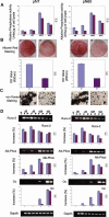
In vitro osteogenic potential of neural crest–derived frontal bone and paraxial mesoderm–derived parietal bone osteoblasts. (A) Alkaline phosphatase activity in pN7 and pN60 frontal and parietal bone osteoblasts. Elevated levels of enzymatic activity were observed in frontal bone osteoblasts. (B) Alizarin red staining and its quantification detected more intense mineralization of extracellular matrix in frontal bone osteoblasts (20× objective magnification). Abbreviations: F = frontal; P = parietal. (C) von Kossa staining revealed the presence of bone nodules of larger size in frontal bone osteoblasts. (D) RT-PCR analysis of osteogenic markers Runx-2, Alp (Alk Phos), and osteocalcin (Oc) in frontal and parietal bone osteoblasts. –rt is a negative control for the reverse transcription reaction performed on pooled RNAs purified from each experimental point. Histogram represents the densitometric analysis of RT-PCR products performed using ImageJ software; the relative intensities of bands were normalized to their respective loading control (Gapdh) and set as 100%; The results are presented as the mean ± SD of three independent experiments.
Fig. 2
Fig. 2
In vivo osteogenic potential of frontal and parietal bones. (A) Two-millimeter calvarial defects were made in the frontal and parietal bones of pN7 mice (upper panel). µCT performed at 2, 4, and 8 weeks after osteotomy. Quantification of bone regeneration (middle panel) (*p ≤ .05). Pentachrome staining (lower panel). (B) Two-millimeter calvarial defects were made in the frontal and parietal bones of pN60 mice (upper panel). Quantification of bone regeneration (middle panel) (*p ≤ .05). Pentachrome staining (lower panel). Arrows indicate the edges of defect. Scale bars = 150 µm in 20× magnification panels, 50 µm in 40× magnification panels.
Fig. 3
Fig. 3
Enhanced activation of canonical Wnt signaling in frontal bone–derived osteoblasts and frontal bone. (A) Western blotting analysis revealed higher levels of nuclear β-catenin in both pN7 and pN60 frontal bone osteoblasts than in parietal bone osteoblasts. Membranes were stripped and reprobed with anti-Lamin B1 antibody to assess for equal loading and transfer of nuclear protein fractions. (B) Pool of active nonphosphorylated β-catenin was assessed on whole-cell lysates using anti-β-catenin antibody, which specifically detects nonphosphorylated active β-catenin. The membranes were stripped and subsequantially incubated with pan-β-catenin antibody and α-tubulin antibody to assess for the total amount of endogenous β-catenin and to control for equal loading and transfer of the samples. (C) Histogram represents the densitometric analysis of electrophoresis bands; the relative intensities of bands were normalized to their respective loading control and set as 100%. The results are presented as the mean ± SD of three independent experiments. (D) Expression level of endogenous β-catenin, cyclin D1, myc, and axin-2 in pN7 frontal and parietal bone osteoblasts. (E) RT-PCR analysis of endogenous β-catenin, cyclin D1, myc, and axin-2 in pN60 frontal and parietal osteoblasts. Histogram represents the densitometric analysis of RT-PCR products performed as above. The results are presented as the mean ± SE of three independent experiments.
Fig. 4
Fig. 4
Enhanced activation of canonical Wnt signaling in neural crest–derived frontal bone. (A) RT-PCR analysis of endogenous β-catenin, cyclin D1, myc, and axin-2 in frontal and parietal bones. Histogram represents the densitometric analysis of RT-PCR products performed as above. (B) X-gal staining of coronal sections of frontal and parietal bones of axin-2lacZ/+ reporter mice showed more intense staining of frontal bone as result of an enhanced activation of canonical Wnt signaling. Boxed areas are enlarged in the lower panels (40×).
Fig. 5
Fig. 5
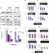
Constitutive activation of canonical Wnt signaling in parietal bone osteoblasts mimics the osteogenic potential of frontal bone osteoblasts. (A) RT-PCR analysis indicated that either in pN7 or pN60 S33Y-infected parietal bone osteoblasts the expression of myc and cyclin D1 genes achieved threshold levels similar to those of control Neo frontal bone osteoblasts. In contrast, expression of Dn-Tcf-4 drastically downregulated the endogenous levels of myc and cyclin D1 in frontal bone osteoblast and parietal bone osteoblasts. (B) Western blotting analysis revealed increased nuclear β-catenin in S33Y-infected parietal bone osteoblasts with levels similar to that of Neo control frontal bone osteoblasts. Decreased accumulation of nuclear β-catenin was observed in both Dn-Tcf-4-infected frontal and parietal bone osteoblasts compared with controls. Filters were stripped and reprobed with anti-Lamin B1 antibody. (C, D) Osteogenic profile determined by alkaline phosphatase activity and alizarin red and von Kossa staining. (E) RT-PCR analysis of osteogenic markers during osteogenic differentiation.
Fig. 6
Fig. 6
Exogenous added Wnt3a confers higher osteogenic potential to parietal bone osteoblasts. (A) Osteogenic differentiation of frontal and parietal bone pN7 and pN60 osteoblasts in the presence or absence of 50 ng/mL Wnt3a assessed by alizarin red staining. (B) Indirect immunofluorescence staining detected intense nuclear staining in Wnt3a-treated parietal bone osteoblasts compared with untreated parietal bone osteoblasts. Increased nuclear translocation of β-catenin also was observed in treated frontal bone osteoblasts, although in a less dramatic manner than in parietal bone osteoblasts. As negative control, a normal primary (irrelevant) mouse IgG was used. Nuclear counterstaining was performed with DAPI (objective magnification 10×). Scale bars = 50 µm.
Fig. 7
Fig. 7
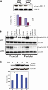
Frontal bone–derived osteoblasts are endowed with higher levels of inactivated GSK-3β than parietal bone–derived osteoblasts. (A) Western blot analysis of cell lysates using a polyclonal phospho-GSK-β3 antibody. Histogram represents the densitometric analysis of electrophoresis bands. (B) Western blot analysis of frontal and parietal bone osteoblasts treated with FGF-2 (20 ng/mL) indicated that FGF-2 treatment increases the pool of phosphorylated GSK-β3. FGF-2-induced phosphorylation of GSK-β3 is abrogated by the presence of Ly294002. Membranes were stripped and reprobed with a monoclonal anti-GSK-β3 antibody to ensure equal loading and transfer of proteins. (C) FGF-2 treatment enhances nuclear translocation of β-catenin. Immunoblotting analysis of nuclear fraction harvested from pN7 parietal bone osteoblasts stimulated with 20 ng/mL of FGF-2 at different time points. One of three independent experiments is shown. Densitometrical analysis revealed increase in nuclear β-catenin, with a peak at 6 hours of stimulation. The results are presented as the mean ± SD of three independent experiments (*p < .03). To show equal protein loading, membranes were stripped and reprobed with anti-Lamin B1 antibody.

References

    1. Morriss-Kay GM. Derivation of the mammalian skull vault. J Anat. 2001;199:143–151. - PMC - PubMed
    1. Jiang X, Iseki S, Maxson RE, Sucov HM, Morriss-Kay GM. Tissue origins and interactions in the mammalian skull vault. Dev Biol. 2002;241:106–116. - PubMed
    1. Chai Y, Jiang X, Ito Y, et al. Fate of the mammalian cranial neural crest during tooth and mandibular morphogenesis. Development. 2000;127:1671–1679. - PubMed
    1. LaBonne C, Bronner-Fraser M. Molecular mechanisms of neural crest formation. Annu Rev Cell Dev Biol. 1999;15:81–112. - PubMed
    1. Anderson DJ. Genes, lineages and the neural crest: a speculative review. Philos Trans R Soc Lond B Biol Sci. 2000;355:953–964. - PMC - PubMed

Publication types

MeSH terms