. 2010 Jan;216(1):3-15.
doi: 10.1111/j.1469-7580.2009.01173.x. Epub 2009 Nov 19.

Developmental stages of the Japanese quail

Affiliations

Developmental stages of the Japanese quail

Sophie J Ainsworth et al. J Anat. 2010 Jan.

Abstract

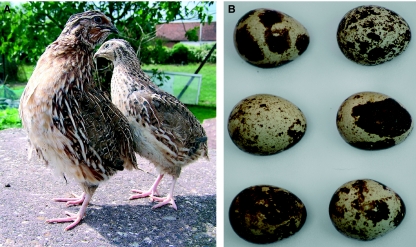
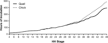
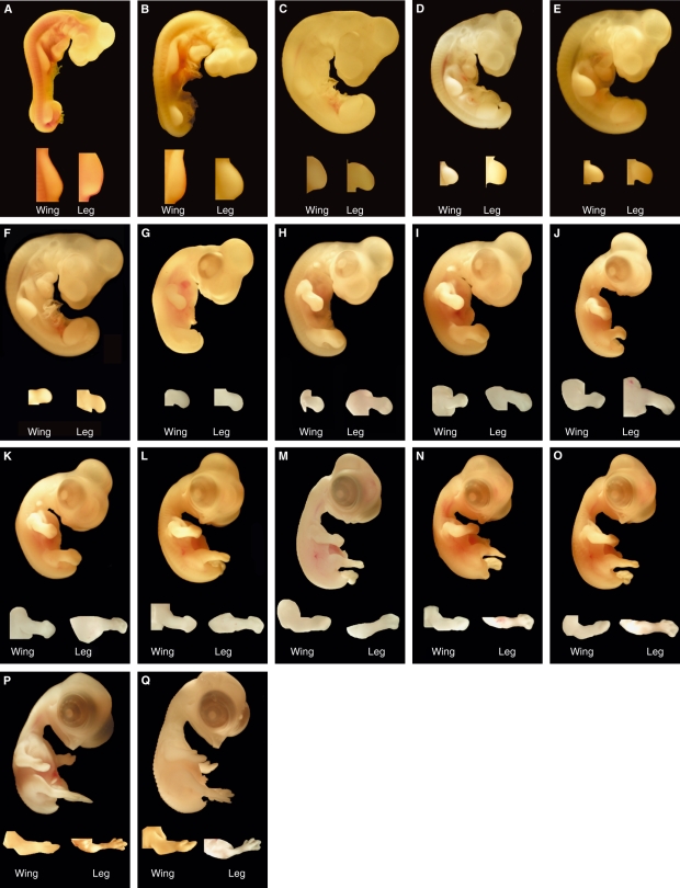
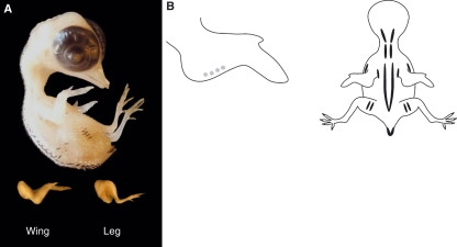
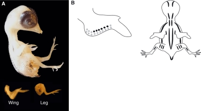
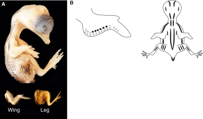
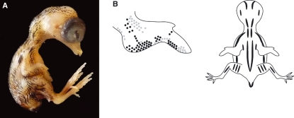
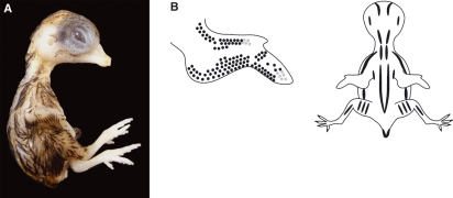
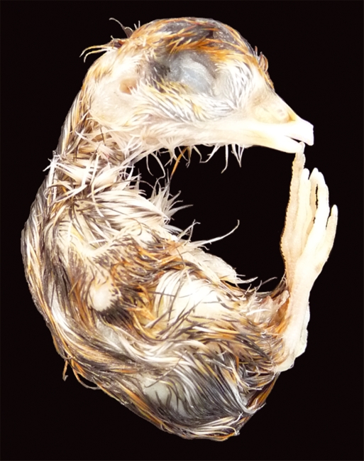
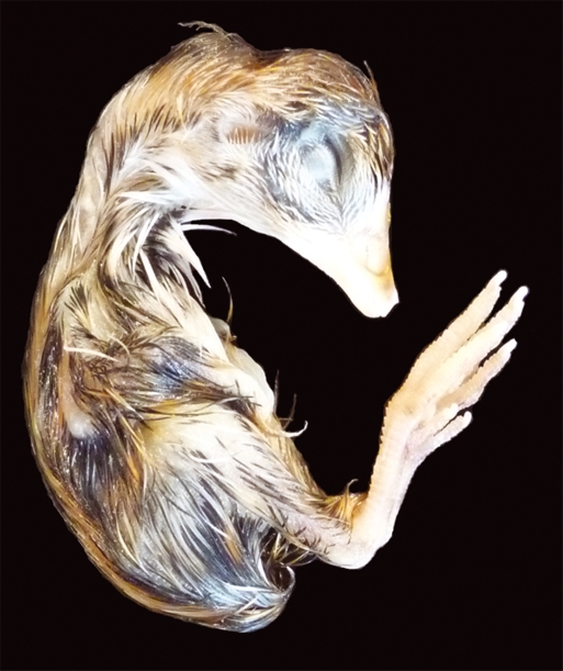
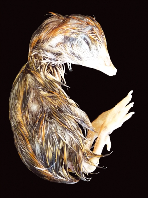
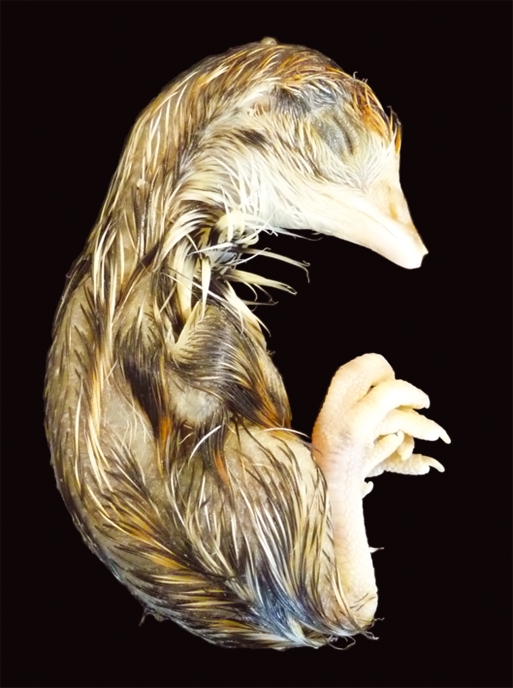
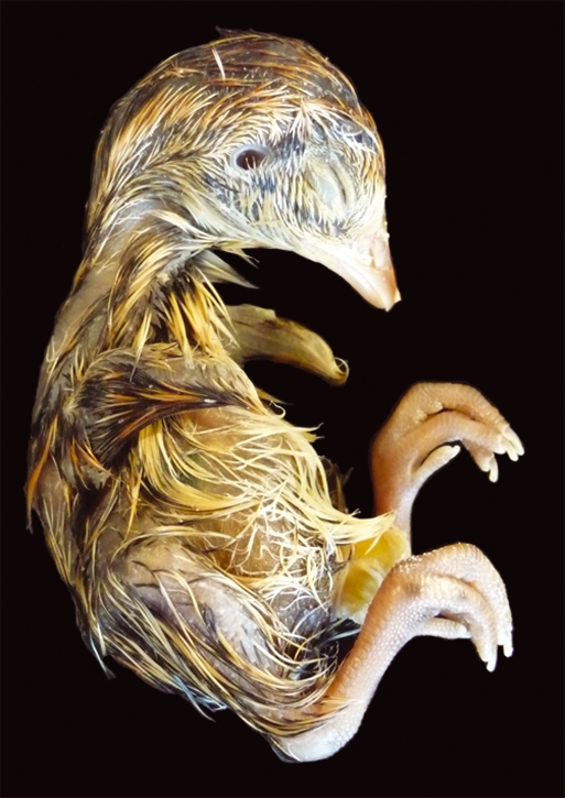
Developmental biology research has used various avian species as model organisms for studying morphogenesis, with the chick embryo being used by the majority of groups. The focus on the chick embryo led Hamburger and Hamilton to develop their definitive staging series nearly 60 years ago and this series is still the mainstay of all laboratories working with avian embryos. The focus on the chick embryo has somewhat overshadowed the importance of another avian embryo that has proved to be equally powerful, the Japanese quail. Since the late 1960s, chimeras have been produced using chick and quail embryos and this technique has revolutionized the approach taken to the investigation of the cellular and molecular interactions that occur during development. Reviews of the literature demonstrate that many research groups are using the quail embryo in a number of established and new ways, and this species has become a primary animal model in developmental biology. Some staging of quail has been performed but this has been incomplete and variations in descriptions, stages and incubation timings mean that comparisons with the chick are not always easily made. There appears to be general agreement that, at the early stages of embryogenesis, there is little developmental difference between chick and quail embryos, although the basis for this has not been established experimentally. The accelerated ontogeny of quail embryos at mid to late stages of development means that registration with the chick is lost. We have therefore developed a definitive developmental stage series for Japanese quail so that differences are fully characterized, misconceptions or assumptions are avoided, and the results of comparative studies are not distorted.


Figures

Fig. 1
Fig. 1
Photographs of adult Japanese quail (A) and a clutch of Japanese quail eggs demonstrating variations in shell colouring (B).
Fig. 2
Fig. 2
Graph showing the average developmental rate (hours of incubation) for Japanese quail and chick embryos, related to HH stages.
Fig. 3
Fig. 3
In-ovo photographs representing quail embryos at stages 4–18, respectively (A–O). Embryos were unfixed, stained with neutral red solution and with vitelline membranes removed.
Fig. 4
Fig. 4
Photographs representing quail embryos, including limb detail, at stages 19–35, respectively (A–Q).
Fig. 5
Fig. 5
(A) Stage 36 (8–9 days) quail embryo, including limb detail. Quail embryos were killed, rinsed and fixed in 5% neutral buffered formalin before photography. (B) Schematic representation of the average pigmentation pattern observed at stage 36 on the wing and the dorsal surface of the embryo. Black and brown pigment is apparent in feather buds. Black pigmentation is present either side of the spine (tapering off in the region of the pelvis), overlying the scapula and ulna, and on the lateral aspects of the thigh and at the edges of the tail. There is a single line of black feathers in the region overlying the coracoids. Golden-brown pigmentation is also present but less extensive, with the most notable pigmentation being in the region of the tail. The beak length (mean length from the anterior angle of the nostril to the tip of the upper bill) is 1.2 mm and the third toe length (mean length from the middle of the metatarsal joint to the end of the most extreme digital pad on the third toe) is 3.2 mm.
Fig. 6
Fig. 6
(A) Stage 37 (9.5 days) quail embryo, including limb detail. (B) Schematic representation of the average pigmentation pattern seen at stage 37 on the wing and the dorsal surface of the embryo. Pigmentation is visible on the head, with black pigmentation present on the forehead and crown. Pigmentation continues to increase either side of the spine over the regions of the scapula and ulna, on the thigh and at the edge of the tail. A single distinct line of black feathers in the region of the coracoids is now prominent, with a second and sometimes third line beginning to appear either side of the first row. Golden-brown pigmentation also increases most notably in the regions of the scapula, ulna, thigh and the edge of the tail. Additional golden-brown pigmentation is apparent in the lumbosacral region when viewed microscopically. The average beak length is 1.5 mm and the third toe length is 4.1 mm.
Fig. 7
Fig. 7
(A) Stage 38 (9.5–10 days) quail embryo, including limb detail. (B) Schematic representation of the average pigmentation pattern seen at stage 38. Black pigmentation is more distinct on the forehead, crown, adjacent to the external auditory meatus, and along the dorsal neck. Pigmentation continues on either side of the spine, over the scapula, ulna, thigh and tail. Several rows of black feathers are visible in the region of the coracoids. Golden-brown pigmentation is now evident in most regions containing black pigmentation and there is a distinct line of golden- brown pigmentation running parallel to the spine. In all regions, mean feather germ lengths have increased. No pigmentation is yet present on the feet. The mean beak length is 1.5 mm and third toe length is 4.7 mm.
Fig. 8
Fig. 8
(A) Stage 39 (10.5–11 days) quail embryo. (B) Schematic representation of the average pigmentation pattern seen at stage 39. Little change in the black pigmentation pattern is observed other than increased pigment appearing on the wing, neck and thorax. Faint pigmentation is also visible for the first time around the intertarsal joint. The length of pigmented feather germs is increased. The mean beak length is 2.0 mm and third toe length is 6.0 mm.
Fig. 9
Fig. 9
(A) Stage 40 (11 days) quail embryo. (B) Schematic representation of the average pigmentation pattern seen at stage 40. Pigmented feather germs are now prominent within the periocular region, particularly ventral to the eye. Increased pigmentation is also visible around the external auditory meatus. Faint pigmentation is evident on the feet around the intertarsal and metatarsal joints. No white feathers can be identified at this stage. The beak length is unchanged at 2.0 mm and third toe length is 6.1 mm.
Fig. 10
Fig. 10
Stage 41 (11.5 days) quail embryo. The length of the feather germs is increased over the whole embryo. White feather germs are apparent throughout the length of the embryo, the most noticeable being ventral to the eye and towards the beak. Pigmentation on the feet is more prominent, particularly around the metatarsal joint. Specific pigmentation patterns are not shown from stage 41 onwards as there is increased variation between embryos. The beak and third toe lengths are unchanged at 2.0 and 6.1 mm, respectively.
Fig. 11
Fig. 11
Stage 42 (12–13 days) quail embryo. Pigmentation on the feet is increasingly prominent and for the first time apparent along the ventral surface of the toes. There is an increased density of feather germs and each feather is considerably longer and produces a ‘glossy’ look. General patterns and features alter little after stage 42 although the embryos continue to increase in size. The overall length of the bird is variable and therefore the beak and third toe length should be used for accurate staging. The mean beak length is 2.3 mm and third toe length is 8.6 mm.
Fig. 12
Fig. 12
Stage 43 (14 days) quail embryo. Mean beak length increases to 2.6 mm and third toe length to 9.4 mm.
Fig. 13
Fig. 13
Stage 44 (15–16 days) quail embryo. Mean beak length is 3.0 mm and third toe length is 10.8 mm.
Fig. 14
Fig. 14
Stage 45 (16–16.5 days) quail embryo. Prehatching stage for the quail embryo with the yolk almost completely internalized, causing the abdomen to swell. Average beak length is 3.5 mm and third toe length is 11.9 mm. Stage 46 (16.5 days) is the hatching stage for the quail embryo.

References

    1. Binder BJ, Landman KA, Simpson MJ, et al. Modeling proliferative tissue growth: a general approach and an avian case study. Phys Rev E Stat Nonlin Soft Matter Phys. 2008;78:031912. - PubMed
    1. Chen Y, Sible JC, McNabb FM. Effects of maternal exposure to ammonium perchlorate on thyroid function and the expression of thyroid-responsive genes in Japanese quail embryos. Gen Comp Endocrinol. 2008;159:196–207. - PubMed
    1. Costa-Silva B, da Costa MC, Melo FR, et al. Fibronectin promotes differentiation of neural crest progenitors endowed with smooth muscle cell potential. Exp Cell Res. 2009;315:955–967. - PubMed
    1. Eames BF, Schneider RA. Quail-duck chimeras reveal spatiotemporal plasticity in molecular and histogenic programs of cranial feather development. Development. 2005;132:1499–1509. - PMC - PubMed
    1. Eames BF, Schneider RA. The genesis of cartilage size and shape during development and evolution. Development. 2008;135:3947–3958. - PMC - PubMed

LinkOut - more resources