. 2011 Feb;15(2):280-95.
doi: 10.1111/j.1582-4934.2009.00973.x.

Role for LAMP-2 in endosomal cholesterol transport

Affiliations

Role for LAMP-2 in endosomal cholesterol transport

Alexander Schneede et al. J Cell Mol Med. 2011 Feb.

Abstract

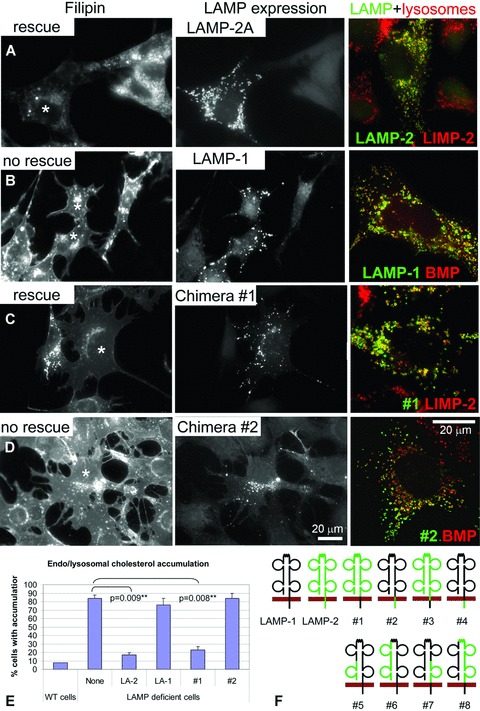
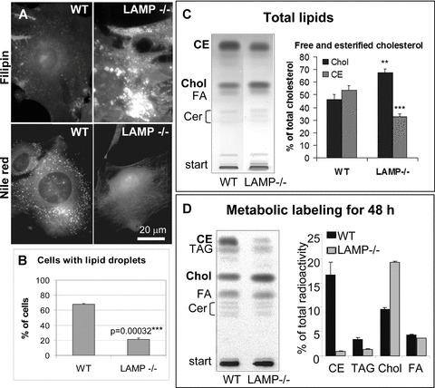
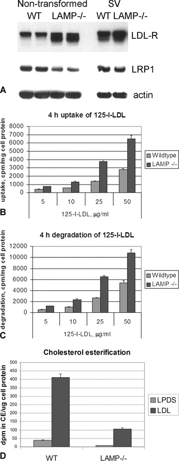
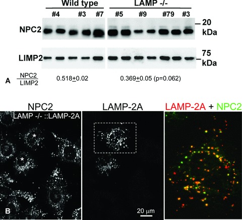
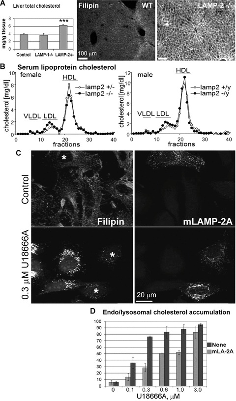
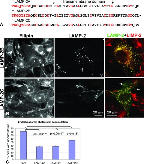
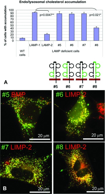
The mechanisms of endosomal and lysosomal cholesterol traffic are still poorly understood. We showed previously that unesterified cholesterol accumulates in the late endosomes and lysosomes of fibroblasts deficient in both lysosome associated membrane protein-2 (LAMP-2) and LAMP-1, two abundant membrane proteins of late endosomes and lysosomes. In this study we show that in cells deficient in both LAMP-1 and LAMP-2 (LAMP(-/-)), low-density lipoprotein (LDL) receptor levels and LDL uptake are increased as compared to wild-type cells. However, there is a defect in esterification of both endogenous and LDL cholesterol. These results suggest that LAMP(-/-) cells have a defect in cholesterol transport to the site of esterification in the endoplasmic reticulum, likely due to defective export of cholesterol out of late endosomes or lysosomes. We also show that cholesterol accumulates in LAMP-2 deficient liver and that overexpression of LAMP-2 retards the lysosomal cholesterol accumulation induced by U18666A. These results point to a critical role for LAMP-2 in endosomal/lysosomal cholesterol export. Moreover, the late endosomal/lysosomal cholesterol accumulation in LAMP(-/-) cells was diminished by overexpression of any of the three isoforms of LAMP-2, but not by LAMP-1. The LAMP-2 luminal domain, the membrane-proximal half in particular, was necessary and sufficient for the rescue effect. Taken together, our results suggest that LAMP-2, its luminal domain in particular, plays a critical role in endosomal cholesterol transport and that this is distinct from the chaperone-mediated autophagy function of LAMP-2.


Figures

Fig 5
Fig 5
Effect of LAMP-2A and LAMP-1 re-expression on late endosomal/lysosomal cholesterol accumulation in LAMP double defi-/ cient MEFs. (A–D) LAMP−/− MEFs were transfected with mouse LAMP-2A (A), rat LAMP-1 (B), chimera #1 (C) or chimera #2 (D), as indicated. One day after transfec-tion, the cells were fixed and stained with filipin to detect cholesterol, and with antimouse LAMP-2 or anti-rat LAMP-1 to detect cells that express the transfected protein. Filipin staining (left panels) and LAMP staining (middle panels) are shown for the same fields. Asterisks indicate the expressing cells in the filipin panels. Immunofluorescence staining was used to study the lysosomal localization of the expressed proteins (right panels in A–D). The samples were double stained for LAMP-2 or LAMP-1 (green) and LIMP-2 or BMP (red). (E) The percentage of / wild-type and LAMP−/− MEFs containing late endosomal/lysosomal cholesterol accumulation was esti-/ mated. The LAMP−/− cells were analysed either without re-expression (None), or when re-expressing LAMP-2A (LA-2), LAMP-1 (LA-1) or one of the LAMP-1/LAMP-2 chimeras #1 or #2. Cells expressing the LAMP protein or chimera were selected for the analysis using LAMP immunofluorescence staining. The results are the mean and S.E.M. from two or three independent experiments. (F) Schematic presentation of the LAMP-1/LAMP-2A chimeras used in this work. Black and green represent LAMP-1 and LAMP-2A, respectively. The red bar represents the lysosomal membrane, with lysosomal lumen above the membrane. The N terminus of LAMP-2 is in the lysosome lumen. The exact location of the boundaries between LAMP-1 and LAMP-2 are presented in Table 1.
Fig 1
Fig 1
Lack of lipid droplets and cholesterol esters in LAMP-1/ LAMP-2 deficient MEFs. (A) Wild-type and LAMP double deficient (LAMP−/−) MEFs were fixed and stained with filipin to detect free cholesterol, or Nile red to detect neutral lipids, as indicated. (B) The percentage of cells with lipid droplets was estimated in wild-type / and LAMP−/− MEFs. (C) Total lipids / of wild-type and LAMP−/− MEFs were analysed using TLC. The quantification of free and esterified cholesterol was done from five experiments and the average and standard deviation are shown. Chol, cholesterol; FA, fatty acids; Cer, ceramide. The asterisks indicate statistical significance: **, P < 0.005; ***, P < 0.001. (D) MEFs were metabolically labelled with [14C] acetate for 48 hrs. Lipids were extracted and same amounts of radioactivity were separated by TLC. The quantification shows mean ± S.E.M. from three experiments. TAG, triacyl glycerol.
Fig 2
Fig 2
Uptake, degradation and esterification of LDL-cholesterol in wild-/ type and LAMP−/− MEFs. (A) The expression levels of LDLR and LRP1 were determined by Western blotting in non-transformed MEFs and in MEFs transformed using the Simian virus large T antigen (SV). β-actin is shown as a loading control. (B) To determine LDL uptake, MEFs were incubated with 5–50 μg/ml [125I]-LDL for 4 hrs at 37°C. Internalized radioactivity was determined. (C) To estimate the degradation of internalized LDL, the content of 125I-tyrosine in the supernatant was determined after 4 hrs incubation. In (B) and (C), the values of specific uptake and degradation represent the mean and S.E.M. of three independent experiments. (D) Cholesterol esterification as a response to LDL loading. Cells grown in lipoprotein deprived medium were pulsed for 6 hrs with [3H] oleic acid in the absence (LPDS) or presence (LDL) of 50 μg/ml LDL. The radioactivity incorporated into CE, per microgram of cell protein, was analysed. The bars represent mean and S.E.M. of two to three individual samples.
Fig 3
Fig 3
Expression and localization of / NPC2 protein in LAMP−/− MEFs. (A) Western blotting of NPC2 in wild-/ type and LAMP−/− MEFs. The numbers above the blots indicate individual cell lines. The lower half of the blot was stained for NPC2 and the upper half was stained for LIMP-2 as a loading control. Our studies have indicated that LIMP-2 is not up-reg-/ ulated in the LAMP−/− MEFs. The signals were quantified and the ratio of NPC2 to LIMP-2 was calculated for each cell line. The averages for each genotype are indicated below the blot. Mean and S.E.M. are given / for the wild-type and LAMP−/− MEF lines. The difference between these two genotypes did not reach statistical significance (P > 0.05). (B) Double labelling of endogenous NPC2 and transiently overexpressed LAMP-2A in LAMP−/− MEFs. Asterisks indicate the LAMP-2 expressing cells in the left panel. Overlay of NPC2 (green) and LAMP-2 (red) for the indicated area is shown in the right panel.
Fig 4
Fig 4
Expression of LAMP-2influ-ences cholesterol levels. (A) LAMP-2 deficient liver accumulates cholesterol. Left panel: Total cholesterol was estimated from five wild-type, three LAMP-1–/–, and three LAMP-2−/− livers. The error bars represent standard deviation. The difference between the control and / LAMP-2−/− is statistically significant (P < 0.001). Middle and right panels: Filipin staining of liver sections from six-month-old wild-type / and LAMP-2−/− mice. (B) Gel filtration analysis of plasma lipoprotein / profiles of LAMP-2−/− mice and heterozygote littermate controls (please note that lamp-2 gene is located in the Y chromosome). Total cholesterol levels in the fractions are shown. Plasma samples from four mice were pooled for each group. Note that both female and male / LAMP-2−/− mice show elevated LDL cholesterol levels. (C, D) Overexpression of LAMP-2A retards the U18666A induced cholesterol accumulation. (C) HeLa cells were transfected with mouse LAMP-2A and grown for 29 hrs. The cells were then treated with increasing concentrations of U18666A for 15 hrs, and processed for filipin staining and immunolabelling with antimouse LAMP-2. Asterisks indicate the LAMP-2A overexpressing cells in the left panels. (D) The percentage of cells with late endosomal/lysosomal cholesterol accumulation was estimated. None, HeLa cells with no overexpression; mLA-2A, HeLa cells overexpressing mouse LAMP-2A. The results are the mean and standard deviation from one experiment with two parallel samples. The experiment was repeated with similar results.
Fig 6
Fig 6
LAMP-2A and LAMP-2B show a similar rescue effect. (A) C-terminal amino acid sequences of mouse LAMP-2 isoforms. All three isoforms have the same N-terminal sequences encoding the luminal domain of the protein. The predicted location of the transmembrane domain, amino acids 380–404, is indicated for LAMP-2A. The unifying nomenclature recommended by Eskelinen et al.[10] is used for the mouse LAMP-2 iso-forms. Red colour indicates identical amino acids in all three isoforms. (B, /C) LAMP−/− MEFs were transfected with LAMP-2A, LAMP-2B or LAMP-2C. After 2 days the cells were processed for filipin staining and LAMP-2 immunofluorescence (left and middle panels in B, respectively). Asterisks indicate the LAMP-2 expressing cells in the filipin panels. The correct targeting of the expressed proteins was monitored by double labelling with anti-LAMP-2 (green) and anti-LIMP-2 (red) (right panels in B). LAMP-2B showed a prominent colocalization with endogenous LIMP-2, while LAMP-2C showed less colocalization and was also detectable on the plasma membrane (arrows). (C) The percentage of cells with late endosomal/lysosomal cholesterol accumulation. The results are the mean and S.E.M. from three (iso-forms A and B) or two (isoform C) independent experiments.
Fig 7
Fig 7
The membrane-proximal half of LAMP-2 luminal domain is critical for cholesterol transport. (A) LAMP−/− MEFs were transfected with one of the constructs #5-#8, as indicated, and stained with filipin and anti-LAMP-2 or anti-LAMP-1 2 days later. Schematic drawings of the chimeras are as in Fig. 5F. The percentage of cells containing late endosomal/lysosomal cholesterol accumulation is shown. The results are mean and S.E.M. of two to three parallel samples in one representative experiment. The experiment was repeated two to three times with similar results. (B) The correct late endosomal/lysosomal localization of the chimeras was checked by immunofuorescence using anti-LAMP-1 or LAMP-2 (green) and a lysoso-mal/late endosomal marker (LIMP-2 or BMP, red).

Similar articles

Cited by

References

    1. Eskelinen EL, Tanaka Y, Saftig P. At the acidic edge: emerging functions for lyso-somal membrane proteins. Trends Cell Biol. 2003;13:137–45. - PubMed
    1. Schröder B, Wrocklage C, Pan C, et al. Integral and associated lysosomal membrane proteins. Traffic. 2007;8:1676–86. - PubMed
    1. Hunziker W, Geuze HJ. Intracellular trafficking of lysosomal membrane proteins. Bioessays. 1996;18:379–89. - PubMed
    1. Fukuda M. Lysosomal membrane glyco-proteins. Structure, biosynthesis, and intracellular trafficking. J Biol Chem. 1991;266:21327–30. - PubMed
    1. Carlsson SR, Roth J, Piller F, et al. Isolation and characterization of human lysosomal membrane glycoproteins, h-lamp-1 and h-lamp-2. Major sialoglyco-proteins carrying polylactosaminoglycan. J Biol Chem. 1988;263:18911–9. - PubMed

Publication types

MeSH terms