

The contribution of Toll-like receptor 2 to the innate recognition of a Leishmania infantum silent information regulator 2 protein
- PMID: 19930041
- PMCID: PMC2792133
- DOI: 10.1111/j.1365-2567.2009.03132.x
The contribution of Toll-like receptor 2 to the innate recognition of a Leishmania infantum silent information regulator 2 protein
Abstract
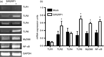
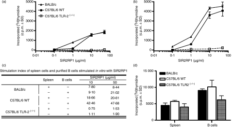
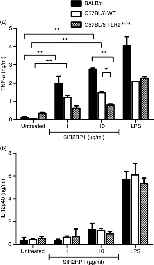
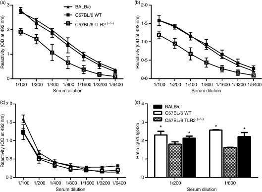
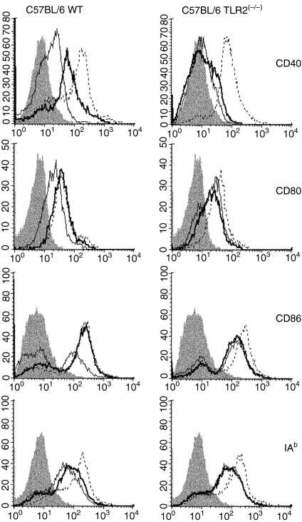
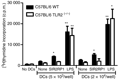
We have characterized a Leishmania protein belonging to the silent information regulator 2 (SIR2) family [SIR2 related protein 1 (SIR2RP1)] that might play an immunoregulatory role during infection through its capacity to trigger B-cell effector functions. We report here that SIR2RP1 leads to the proliferation of activated B cells, causing increased expression of major histocompatibility complex (MHC) II and the costimulatory molecules CD40 and CD86, which are critical ligands for T-cell cross-talk during the development of adaptive immune responses. In contrast, B cells isolated from Toll-like receptor 2 (TLR2) knockout mice were unable to respond to the SIR2RP1 stimulus. Similarly, SIR2RP1 induced the maturation of dendritic cells (DCs) in a TLR2-dependent manner with the secretion of pro-inflammatory cytokines [interleukin (IL)-12 and tumour necrosis factor (TNF)-alpha] and enhanced the costimulatory properties of DCs. Nevertheless, immunization assays demonstrated that TLR2-deficient mice were able to mount a specific humoral response to SIR2RP1. Interestingly, further investigations showed that macrophages were activated by SIR2RP1 even in the absence of TLR2. Therefore, a different type of interplay between SIR2RP1 and the major antigen-presenting cells in vivo could explain the immune response observed in TLR2-deficient mice. Together, these results demonstrate that TLR2 signalling contributes to SIR2RP1 recognition by innate immune host cells.
Figures

Similar articles
-
TLR2- and 4-independent immunomodulatory effect of high molecular weight components from Ascaris suum.Mol Immunol. 2014 Mar;58(1):17-26. doi: 10.1016/j.molimm.2013.10.011. Epub 2013 Nov 19. Mol Immunol. 2014. PMID: 24263181
-
Toll-like receptor-induced innate immune responses in non-parenchymal liver cells are cell type-specific.Immunology. 2010 Mar;129(3):363-74. doi: 10.1111/j.1365-2567.2009.03179.x. Epub 2009 Nov 16. Immunology. 2010. PMID: 19922426 Free PMC article.
-
Contribution of a Streptococcus mutans antigen expressed by a Salmonella vector vaccine in dendritic cell activation.Infect Immun. 2011 Sep;79(9):3792-800. doi: 10.1128/IAI.05338-11. Epub 2011 Jul 11. Infect Immun. 2011. PMID: 21746857 Free PMC article.
-
Leishmania species-dependent functional duality of toll-like receptor 2.IUBMB Life. 2019 Nov;71(11):1685-1700. doi: 10.1002/iub.2129. Epub 2019 Jul 22. IUBMB Life. 2019. PMID: 31329370 Review.
-
Paradoxical immune response in leishmaniasis: The role of toll-like receptors in disease progression.Parasite Immunol. 2022 Apr;44(4-5):e12910. doi: 10.1111/pim.12910. Epub 2022 Feb 28. Parasite Immunol. 2022. PMID: 35119120 Free PMC article. Review.
Cited by
-
Deception and manipulation: the arms of leishmania, a successful parasite.Front Immunol. 2014 Oct 20;5:480. doi: 10.3389/fimmu.2014.00480. eCollection 2014. Front Immunol. 2014. PMID: 25368612 Free PMC article. Review.
-
Expression of toll-like receptor 2, 4 and related cytokines in intraperitoneally inoculated Balb/C mice with Echinococcus multilocularis.Int J Clin Exp Pathol. 2017 Jul 1;10(7):7947-7955. eCollection 2017. Int J Clin Exp Pathol. 2017. PMID: 31966645 Free PMC article.
-
The type III histone deacetylase Sirt1 protein suppresses p300-mediated histone H3 lysine 56 acetylation at Bclaf1 promoter to inhibit T cell activation.J Biol Chem. 2011 May 13;286(19):16967-75. doi: 10.1074/jbc.M111.218206. Epub 2011 Mar 22. J Biol Chem. 2011. PMID: 21454709 Free PMC article.
-
Toll receptors type-2 and CR3 expression of canine monocytes and its correlation with immunohistochemistry and xenodiagnosis in visceral leishmaniasis.PLoS One. 2011;6(11):e27679. doi: 10.1371/journal.pone.0027679. Epub 2011 Nov 30. PLoS One. 2011. PMID: 22140456 Free PMC article.
-
More than just exosomes: distinct Leishmania infantum extracellular products potentiate the establishment of infection.J Extracell Vesicles. 2018 Nov 8;8(1):1541708. doi: 10.1080/20013078.2018.1541708. eCollection 2019. J Extracell Vesicles. 2018. PMID: 30455859 Free PMC article.
References
-
- Medzhitov R, Janeway CA., Jr Innate immunity: impact on the adaptive immune response. Curr Opin Immunol. 1997;9:4–9. - PubMed
-
- Muzio M, Mantovani A. Toll-like receptors. Microbes Infect. 2000;2:251–5. - PubMed
-
- Janeway CA, Jr, Medzhitov R. Innate immune recognition. Annu Rev Immunol. 2002;20:197–216. - PubMed
-
- Kawai T, Akira S. TLR signaling. Semin Immunol. 2007;19:24–32. - PubMed
-
- Carpenter S, O’Neill LA. How important are Toll-like receptors for antimicrobial responses? Cell Microbiol. 2007;9:1891–901. - PubMed
Publication types
MeSH terms
Substances
LinkOut - more resources
Full Text Sources
Molecular Biology Databases
Research Materials
