. 2009 Dec;215(6):611-35.
doi: 10.1111/j.1469-7580.2009.01156.x.

The phylogeny of the red panda (Ailurus fulgens): evidence from the forelimb

Affiliations

The phylogeny of the red panda (Ailurus fulgens): evidence from the forelimb

Rebecca E Fisher et al. J Anat. 2009 Dec.

Abstract

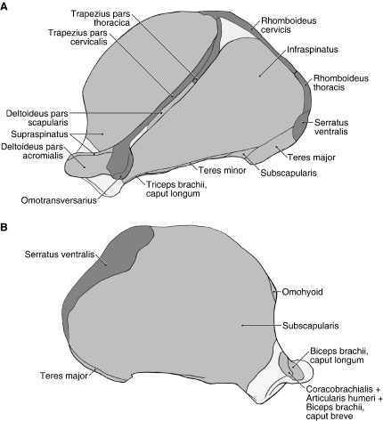
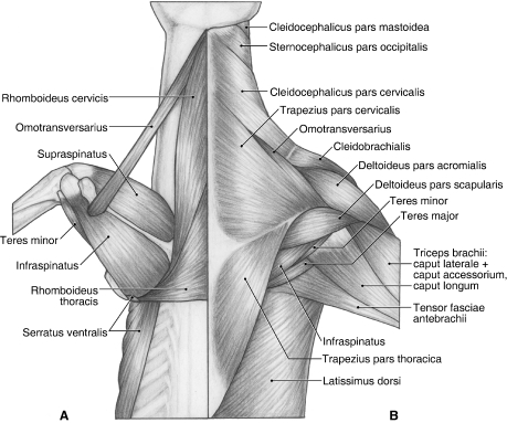
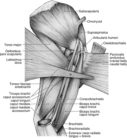
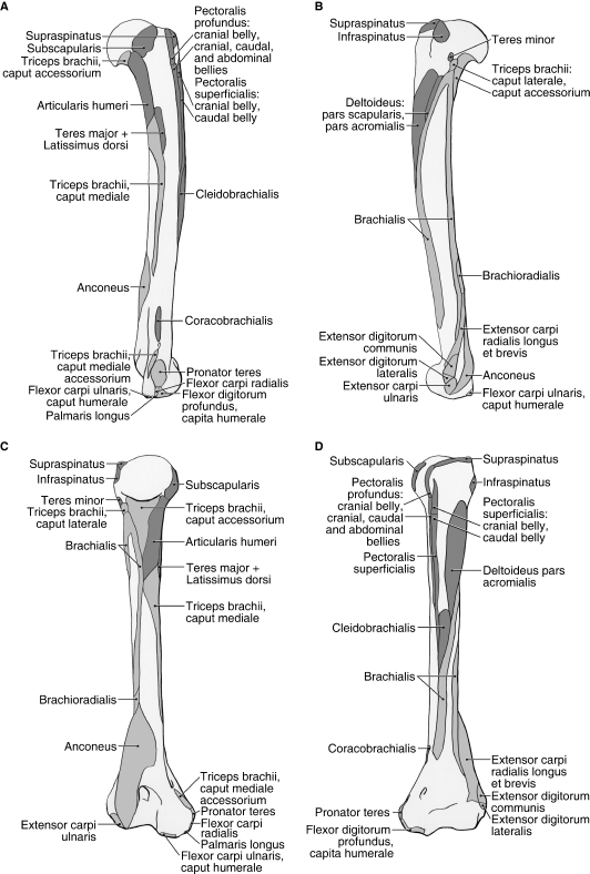
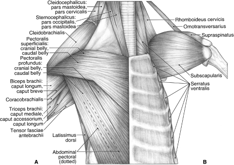
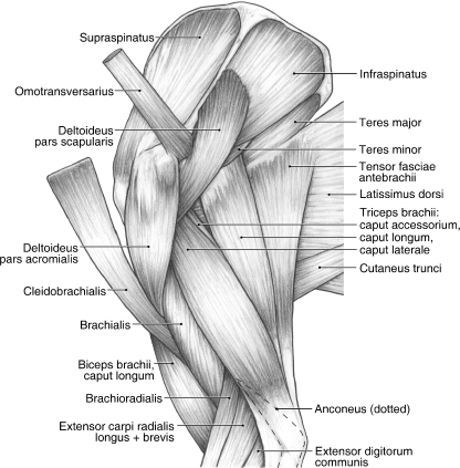
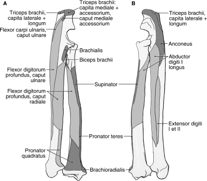
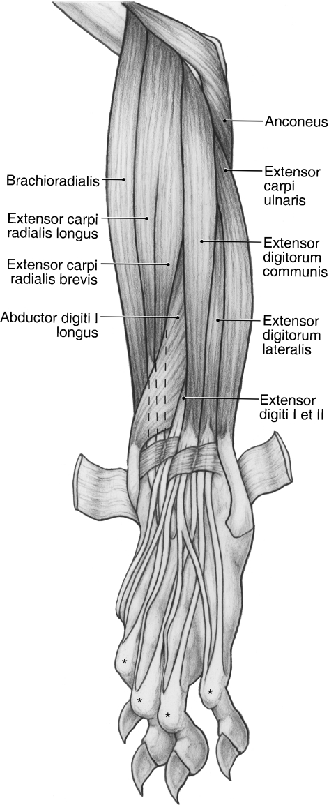
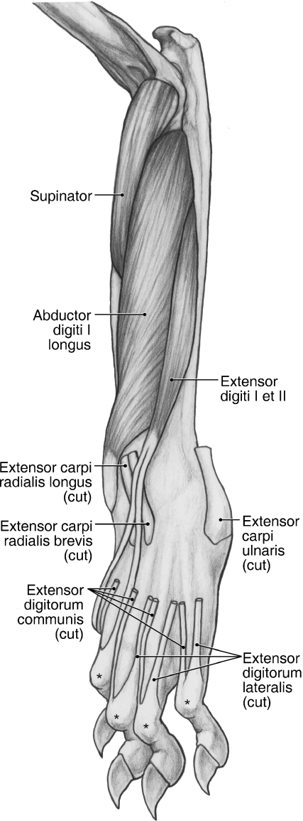
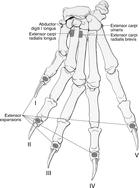
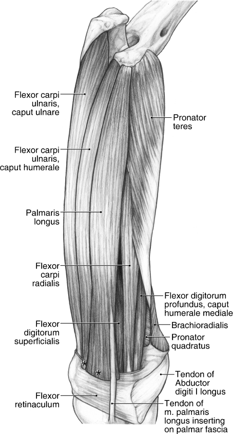
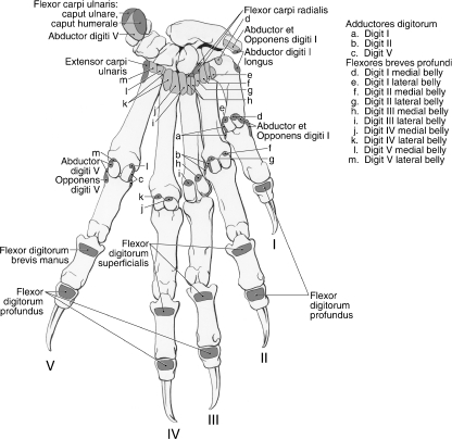
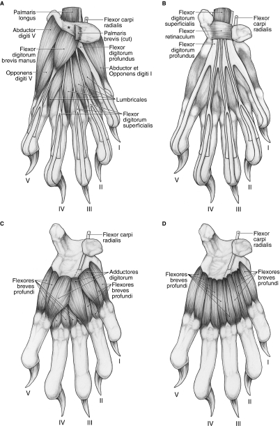
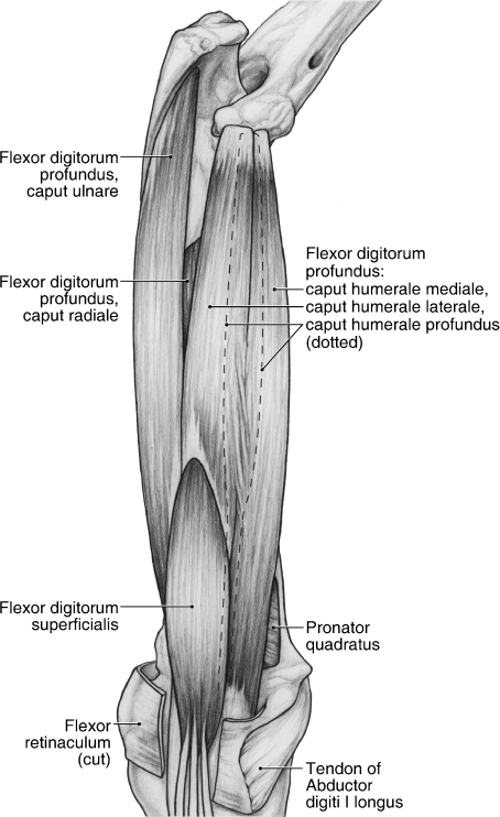
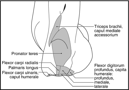
Within the order Carnivora, the phylogeny of the red panda (Ailurus fulgens) is contentious, with morphological and molecular studies supporting a wide range of possible relationships, including close ties to procyonids, ursids, mustelids and mephitids. This study provides additional morphological data, including muscle maps, for the forelimb of Ailurus, based on the dissection of four cadavers from the National Zoological Park, Washington, DC, USA. The red panda forelimb is characterized by a number of primitive features, including the lack of m. rhomboideus profundus, a humeral insertion for m. cleidobrachialis, the presence of mm. brachioradialis, articularis humeri and coracobrachialis, a single muscle belly for m. extensor digitorum lateralis with tendons to digits III-V, four mm. lumbricales, and the presence of mm. flexor digitorum brevis manus, adductores digiti I, II and V, and abductor digiti I and V. Red pandas resemble Ailuropoda, mustelids and some procyonids in possessing a soft tissue origin of m. flexor digitorum superficialis. In addition, red pandas are similar to ursids and procyonids in having a variable presence of m. biceps brachii caput breve. Furthermore, Ailurus and some ursids lack m. rhomboideus capitis. The forelimb muscle maps from this study represent a valuable resource for analyzing the functional anatomy of fossil ailurids and some notes on the Miocene ailurid, Simocyon batalleri, are presented.


Figures

Fig. 2
Fig. 2
Scapula muscle maps for Ailurus (left side), including the origin of m. deltoideus pars scapularis from the scapular spine (present in four limbs). (A) Lateral view; (B) medial view.
Fig. 1
Fig. 1
Dorsal view of the back and shoulder in Ailurus. (A) Deep view; (B) superficial view.
Fig. 4
Fig. 4
Medial view of the left shoulder and brachium in Ailurus, including m. biceps brachii caput breve (present in four limbs). *Fibers of m. latissimus dorsi coursing superficially to join m. pectoralis profundus.
Fig. 6
Fig. 6
Humerus muscle maps for Ailurus (left side), including insertions of the cranial and caudal bellies of m. pectoralis superficialis (present in six limbs). (A) medial view; (B) lateral view; (C) caudal view; (D) cranial view.
Fig. 3
Fig. 3
Ventral view of the chest and shoulder in Ailurus, including m. biceps brachii caput breve (present in four limbs) and a small, partially separable cranial belly of m. pectoralis superficialis (present in six limbs). (A) Superficial view; (B) deep view.
Fig. 5
Fig. 5
Lateral view of the left shoulder and brachium in Ailurus, including the origin of m. deltoideus pars scapularis from the scapular spine (present in four limbs).
Fig. 7
Fig. 7
Radius and ulna muscle maps for Ailurus (left side). (A) Caudomedial view; (B) craniolateral view.
Fig. 8
Fig. 8
Superficial dissection of the craniolateral antebrachium and dorsal manus in the left forelimb of Ailurus. *The extensor expansions.
Fig. 10
Fig. 10
Deep dissection of the craniolateral antebrachium and dorsal manus in the left forelimb of Ailurus. *The extensor expansions.
Fig. 9
Fig. 9
Dorsal manus muscle map for Ailurus (left side).
Fig. 11
Fig. 11
Superficial dissection of the caudomedial antebrachium of the left forelimb in Ailurus, including fusion of m. pronator teres in its proximal two-thirds with m. flexor carpi radialis (present in six limbs) and separable mm. flexor carpi ulnaris capita humerale et ulnare (present in seven limbs). *The cut edge of mm. flexor carpi ulnaris caput humerale (present in four limbs) and palmaris longus where they insert onto the superficial palmar fascia.
Fig. 13
Fig. 13
Palmar manus muscle map for Ailurus, including the separate insertions of mm. flexor carpi ulnaris capita humerale et ulnare (present in seven limbs) (left side).
Fig. 15
Fig. 15
Palmar views of the manus in Ailurus (left side). (A) First (superficial) layer; (B) second layer; (C) third layer; (D) fourth (deep) layer, including two subdivisions in the medial belly of m. flexor brevis profundus digiti III (present in six limbs). *The superficial connective tissue spanning the pisiform and radial sesamoid.
Fig. 14
Fig. 14
Deep dissection of the caudomedial antebrachium of the left forelimb in Ailurus.
Fig. 12
Fig. 12
Close-up view of the left medial epicondyle muscle map in Ailurus.

References

    1. Allen H. The muscles of the limbs of the raccoon (Procyon lotor) Proc Acad Nat Sci Philadelphia. 1882:115–144.
    1. Antón M, Salesa MJ, Pastor J, et al. Implications of the functional anatomy of the hand and forearm of Ailurus fulgens (Carnivora, Ailuridae) for the evolution of the ‘false-thumb’ in pandas. J Anat. 2006;209:757–764. - PMC - PubMed
    1. Beddard FE. On the visceral and muscular anatomy of Cryptoprocta ferox. Proc Zool Soc Lond. 1895:430–437.
    1. Beddard FE, Treves F. On the anatomy of Rhinoceros sumatrensis. Proc Zool Soc Lond. 1889;1:7–25.
    1. Beswick-Perrin J. On the myology of the limbs of the Kinkajou (Cercoleptes caudivolvulus) Proc Zool Soc Lond. 1871:547–559.

Publication types

LinkOut - more resources