. 2009;10(11):R135.
doi: 10.1186/gb-2009-10-11-r135. Epub 2009 Nov 24.

Expansion of the human mitochondrial proteome by intra- and inter-compartmental protein duplication

Affiliations

Expansion of the human mitochondrial proteome by intra- and inter-compartmental protein duplication

Radek Szklarczyk et al. Genome Biol. 2009.

Abstract

Background: Mitochondria are highly complex, membrane-enclosed organelles that are essential to the eukaryotic cell. The experimental elucidation of organellar proteomes combined with the sequencing of complete genomes allows us to trace the evolution of the mitochondrial proteome.

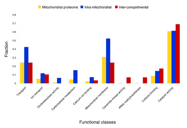
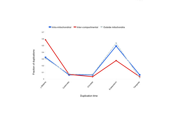
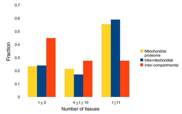
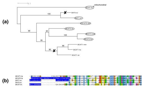
Results: We present a systematic analysis of the evolution of mitochondria via gene duplication in the human lineage. The most common duplications are intra-mitochondrial, in which the ancestral gene and the daughter genes encode mitochondrial proteins. These duplications significantly expanded carbohydrate metabolism, the protein import machinery and the calcium regulation of mitochondrial activity. The second most prevalent duplication, inter-compartmental, extended the catalytic as well as the RNA processing repertoire by the novel mitochondrial localization of the protein encoded by one of the daughter genes. Evaluation of the phylogenetic distribution of N-terminal targeting signals suggests a prompt gain of the novel localization after inter-compartmental duplication. Relocalized duplicates are more often expressed in a tissue-specific manner relative to intra-mitochondrial duplicates and mitochondrial proteins in general. In a number of cases, inter-compartmental duplications can be observed in parallel in yeast and human lineages leading to the convergent evolution of subcellular compartments.

Conclusions: One-to-one human-yeast orthologs are typically restricted to their ancestral subcellular localization. Gene duplication relaxes this constraint on the cellular location, allowing nascent proteins to be relocalized to other compartments. We estimate that the mitochondrial proteome expanded at least 50% since the common ancestor of human and yeast.


Figures

Figure 1
Figure 1
Contribution of different duplication types to the function of the mitochondria. Classes significantly overrepresented compared to the mitochondrial proteome are shown. The height of a bar represents the fraction of proteins that is annotated with a specific category. Three datasets are shown: the whole mitochondrial proteome (MitoCarta proteins [4]; yellow), intra-mitochondrial (blue) and inter-compartmental (red) duplications. Protein functional classes are defined by GO functional classification [68]. Benjamini and Hochberg false discovery rate correction was used to derive statistically significant categories. See Tables S5 and S7 in Additional data file 1 for the full list.
Figure 2
Figure 2
Timing of gene duplications of mitochondrial proteins. The solid blue line represents duplicating mitochondrial proteins, while the solid red line corresponds to duplications of genes followed by relocalization of one of the proteins to the mitochondria. The dashed line denotes protein duplications in other cellular compartments, outside the mitochondria (all proteins are listed in Table S9 in Additional data file 1).
Figure 3
Figure 3
Expression profiles of mitochondrial proteins across a range of mouse tissues. The number of tissues with a detectable protein mass-spectrometry signal (up to 14 tissues investigated in [4]) is shown. The height of the bar represents the fraction of proteins - of intra-mitochondrial or inter-compartmental duplication origins - expressed in a tissue-specific manner (up to three tissues), widely expressed (in more than ten tissues) or expressed in a moderate number of tissues. Figure S2 in Additional data file 1 presents the data in more detail.
Figure 4
Figure 4
Evolution of mitochondrial localization for the branched-chain-amino-acid aminotransferases family. (a) Gene tree generated by PhyML [70] for vertebrates, yeast and outgroup species that speciated before duplication events. Bootstrap values (100 repetitions) are shown on the internal branches. Proteins surrounded by an oval are localized to mitochondria; loss of the mitochondrial localization is marked by a cross. (b) Clustal W [71] alignment of the amino-terminal region of orthologs. The predicted targeting sequences are highlighted in blue. Abbreviations: hs. Homo sapiens; mm, Mus musculus; dm, Drosophila melanogaster; dr, Dano rerio; lm, L. major; sc, S. cerevisiae.

References

    1. Green DR, Reed JC. Mitochondria and apoptosis. Science. 1998;281:1309–1312. doi: 10.1126/science.281.5381.1309. - DOI - PubMed
    1. Berridge MJ, Lipp P, Bootman MD. The versatility and universality of calcium signalling. Nat Rev Mol Cell Biol. 2000;1:11–21. doi: 10.1038/35036035. - DOI - PubMed
    1. Meisinger C, Sickmann A, Pfanner N. The mitochondrial proteome: from inventory to function. Cell. 2008;134:22–24. doi: 10.1016/j.cell.2008.06.043. - DOI - PubMed
    1. Pagliarini DJ, Calvo SE, Chang B, Sheth SA, Vafai SB, Ong S, Walford GA, Sugiana C, Boneh A, Chen WK, Hill DE, Vidal M, Evans JG, Thorburn DR, Carr SA, Mootha VK. A mitochondrial protein compendium elucidates complex I disease biology. Cell. 2008;134:112–123. doi: 10.1016/j.cell.2008.06.016. - DOI - PMC - PubMed
    1. Karlberg O, Canbäck B, Kurland CG, Andersson SG. The dual origin of the yeast mitochondrial proteome. Yeast. 2000;17:170–187. doi: 10.1002/1097-0061(20000930)17:3<170::AID-YEA25>3.0.CO;2-V. - DOI - PMC - PubMed

LinkOut - more resources