. 2009 Nov 24;16(1):105.
doi: 10.1186/1423-0127-16-105.

Molecular events associated with epithelial to mesenchymal transition of nasopharyngeal carcinoma cells in the absence of Epstein-Barr virus genome

Affiliations

Molecular events associated with epithelial to mesenchymal transition of nasopharyngeal carcinoma cells in the absence of Epstein-Barr virus genome

Jung-Chung Lin et al. J Biomed Sci. .

Abstract

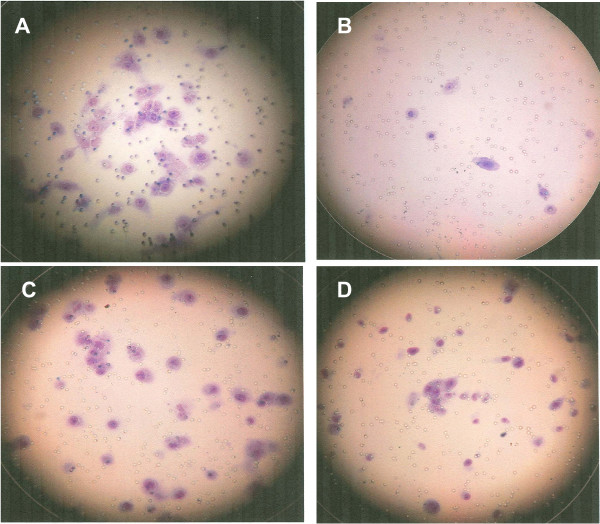
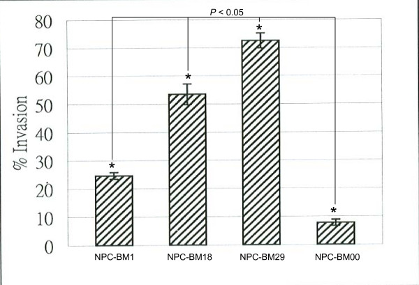
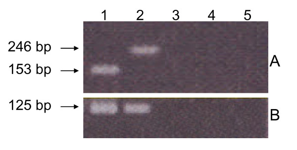
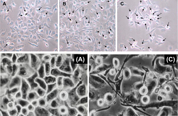
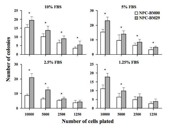
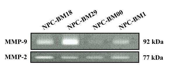
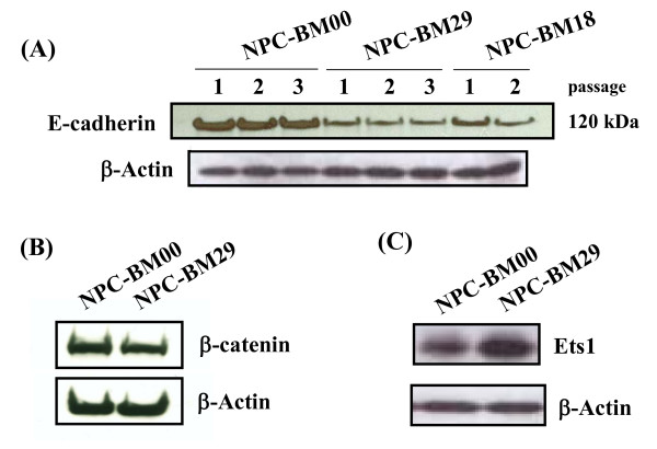
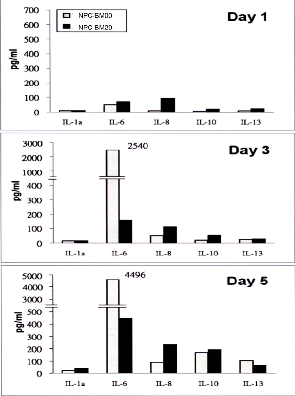
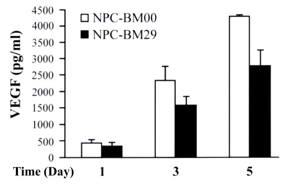
Epithelial-mesenchymal transition (EMT) is an important process in tumor metastasis. The EMT-related events associated with metastasis of NPC in the absence of EBV have not been elucidated. We established an EBV-negative NPC cell line from a bone marrow biopsy of an NPC patient. Using a Matrigel system we isolated an invasive and non-invasive sublines, designated NPC-BM29 and NPC-BM00. NPC-BM29 acquired an invasive-like phenotype characterized by EMT, marked by down-regulation of E-cadherin and beta-catenin with concomitant increased expression of Ets1. NPC-BM29 cells expressed >or= 10-fold higher of MMP-9 than NPC-BM00 cells. NPC-BM29 cells grew better in 2% serum than NPC-BM00 cells, with a population doubling-time of 26.8 h and 30.7 h, respectively. A marked reduction in colony-formation ability of NPC-BM00 cells compared to NPC-BM29 was observed. Wound-healing assay revealed that NPC-BM29 cells displayed higher motility than NPC-BM00 and the motility was further enhanced by cell treatment with TPA, a PKC activator. Cell surface markers and tumor-associated molecules, AE3, MAK6 and sialyl-Tn, were up-regulated in NPC-BM29 cells, whereas the expression of HLA-DR and CD54 was significantly increased in NPC-BM00 cells. NPC-BM29 consistently released higher levels of IL-8 and IL-10 than NPC-BM00, with low levels of IL-1alpha expression in both cell lines. Higher level of VEGF production was detected in NPC-BM00 than NPC-BM29 cells. These data show that EBV is not required for exhibiting multiple metastatic phenotypes associated with EMT. More studies that target right molecules/signalings associated with the EMT may offer new therapeutic intervention options for NPC invasion and metastasis.


Figures

Figure 1
Figure 1
Selection of invasive cells by Matrigel Invasion Chambers. NPC-BM1 cells were seeded onto the filters which were coated with the reconstituted Matrigel of the upper compartment of each chamber and incubated with DMEM. After incubation, cells on the upper side of the filters were removed and cells in the lower surface of the filter were stained with Diff-Quick solution for counting. A, invasive cells (NPC-BM18) after the first round of selection; B, non-invasive cells (NPC-BM00); C, invasive cells (NPC-BM29) after the second round of selection; D, the parental cells (NPC-BM1).
Figure 2
Figure 2
Quantitation of migrated cells. Cells (1 × 10⁴) were plated on the Matrigel. After 48 h, the invasive cells were stained with Diff-Quick solution. The number of invasive cells was counted from three different fields.
Figure 3
Figure 3
Detection of EBV DNA by polymerase chain reaction. Panel A, a fragment of 153 bp and 246 bp of EBNA-3C were detected in B95-8 (lane 1) and AG876 (lane 2); NPC-BM1 (lane 3), NPC-BM00 (lane 4) and NPC-BM29 (lane 5) were all negative for EBNA-3C. Panel B, an expected fragment of 125 bp of BamHI W were detected in B95-8 (lane 1) and AG876 (lane 2), but not in NPC-BM1, NPC-BM00 and NPC-BM29.
Figure 4
Figure 4
Morphological changes of NPC-BM1 cells after separation of invasive from non-invasive cells. Cells were plated in low density and photographed after 36 h using light microscopy (× 200 magnification). Panel A, parental cells (NPC-BM1); B, cells (NPC-BM18) after the first round of selection; C, cells (NPC-BM29) after the second round of selection. NPC BM1 exhibited typical well-attached polygonal epithelial cell morphology with few round and packed appearance cells (arrows) representing EMT morphology. These EMT cells increased in NPC-BM18 and NPC-BM29. The epithelial to mesenchymal transition (EMT) is characterized by the loss of epithelial characteristics and the gain of mesenchymal attributes in epithelial cells. Panels (A) and (C) are the higher power view of panels A and C. Magnification 450 ×.
Figure 5
Figure 5
Effect of low-serum medium on cell proliferation of NPC-BM00 and NPC-BM29 cells. To compare the growth rate, both NPC-BM00 and NPC-BM29 were seeded and cultured in 2% FBS. The cell proliferation was monitored daily for 8 days. NPC-BM29 grew better in low serum medium than NPC-BM00.
Figure 6
Figure 6
Anchorage-independent cell growth by soft agar assay. Soft agar assay was used to compare the colony forming ability of NPC-BM00 and NPC-BM29 cells in medium containing different concentrations of fetal calf serum (FBS) and cell numbers. Both the NPC-BM00 and NPC-BM29 cells were seeded with 1250, 2500, 5000 and 10000 cells per well in 24-well plates. The colonies were counted after 6 days of culture. Data are presented as the means ± standard deviation from experiments that were performed in triplicate wells. * referred to the level of significance at P < .05 and # P > .05.
Figure 7
Figure 7
Scrape-wound migration assay. Confluent monolayers of NPC-BM00 and NPC-BM29 were grown in the presence and absence of tumor promoting agent TPA and were scraped by a plastic pipette. The wound-induced cell motility was observed after 7 h. Panel A, NPC-BM00 and NPC-BM29 cells in the presence and absence of TPA at 0 h after scraping; Panel B, cell motility 7 h after scraping. Magnification, 150 ×.
Figure 8
Figure 8
Differential expression of MMP-9 by the NPC-BM1 parental line and its sublines (NPC-BM00. --BM18, -BM280) detected by gelatin zymography. Cells were cultured for 3 days and the condition media were harvested and processed for gelatin zymography analysis as described in Materials and Methods. Lane 1, conditioned medium from NPC-BM18; lane 2, from NPC-BM29; lane 3, from NPC-BM00; lane 4, from parental cell NPC-BM1.
Figure 9
Figure 9
Down-regulation of intracellular adhesion molecules E-cadherin and -catenin in NPC-BM clones. E-cadherin and β-catenin in cell lysates of NPC-BM00, NPC-BM29 and NPC-BM18 were analyzed by Western blot using monoclonal antibodies specific for these molecules. Specific Western blot signals were visualized on an X-ray film by incubating with ECL-Plus chemiluminescence reagents. Panel A, E-cadherin; panel B, -catenin. The expression of E-cadherin remained unchanged in cells of different passages. Upregulation of transcriptional factor Ets1 was detected in NPC-BM29 cells (panel C). Both NPC-BM00 and NPC-BM29 cells were processed for Western blot analysis the same way as done for E-cadherin and -catenin except the monoclonal antibody specific for Ets1 was used.
Figure 10
Figure 10
The cytokine expression profiles of NPC-BM00 and NPC-BM29 cells. The cultured media from NPC-BM00 and NPC-BM29 cells were collected and analyzed by the Q-Plex™ Human Cytokine Array for a panel of 12 distinct cytokines as described in the text. The cytokine expression profiles were monitored and analyzed from cultured media collected on day 1, 3, and 5.
Figure 11
Figure 11
Detection of VEGF production by ELISA. The cultured media from NPC-BM00 and NPC-BM29 cells were collected and analyzed by ELISA using VEGF monoclonal antibody coated on the microtiter plate. The VEGF expression profiles were monitored and analyzed from cultured media collected on day 1, 3, and 5.

References

    1. Rickinson AB, Kieff E. In: Fields Virology. 3. Fields BN, Knipe DM, Howley PM, editor. Philadelphia; Lippincott-Raven; 1996. Epstein-Barr virus; pp. 2391–2246.
    1. Lin JC. In: New Developments in Epstein-Barr Virus. Umar CS, editor. New York; Nova Science Publishers, Inc; 2006. Pathogenesis and therapy of Epstein-Barr virus infection: Novel therapeutic approaches; pp. 1–35.
    1. Ortiz-McWilliams JO. In: Head and Neck Oncology. 1. Myers EM, editor. Boston; Little-Brown; 1991. History and physical examination; pp. 19–43.
    1. Pagano JS, Blaser M, Buendia MA, Damania B, Khalili K, Raab-Traub N, Roizman B. Infectious agents and cancer: criteria for a causal relation. Semin Cancer Biol. 2004;14:453–71. - PubMed
    1. Rosenthal EL, Matrisian LM. Matrix metalloproteases in head and neck cancer. Head Neck. 2006;28:639–48. - PMC - PubMed

Publication types