. 2009 Nov 24:8:110.
doi: 10.1186/1476-4598-8-110.

PRL-3 promotes the motility, invasion, and metastasis of LoVo colon cancer cells through PRL-3-integrin beta1-ERK1/2 and-MMP2 signaling

Affiliations

PRL-3 promotes the motility, invasion, and metastasis of LoVo colon cancer cells through PRL-3-integrin beta1-ERK1/2 and-MMP2 signaling

Lirong Peng et al. Mol Cancer. .

Abstract

Background: Phosphatase of regenerating liver-3 (PRL-3) plays a causative role in tumor metastasis, but the underlying mechanisms are not well understood. In our previous study, we observed that PRL-3 could decrease tyrosine phosphorylation of integrin beta1 and enhance activation of ERK1/2 in HEK293 cells. Herein we aim to explore the association of PRL-3 with integrin beta1 signaling and its functional implications in motility, invasion, and metastasis of colon cancer cell LoVo.

Methods: Transwell chamber assay and nude mouse model were used to study motility and invasion, and metastsis of LoVo colon cancer cells, respectively. Knockdown of integrin beta1 by siRNA or lentivirus were detected with Western blot and RT-PCR. The effect of PRL-3 on integrin beta1, ERK1/2, and MMPs that mediate motility, invasion, and metastasis were measured by Western blot, immunofluorencence, co-immunoprecipitation and zymographic assays.

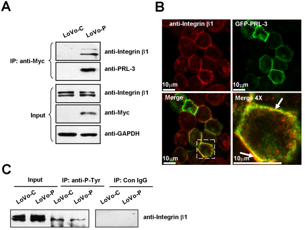
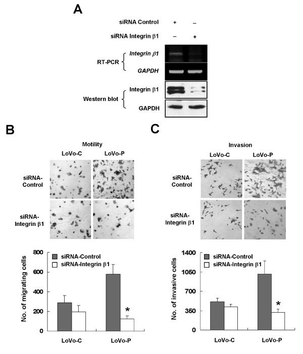
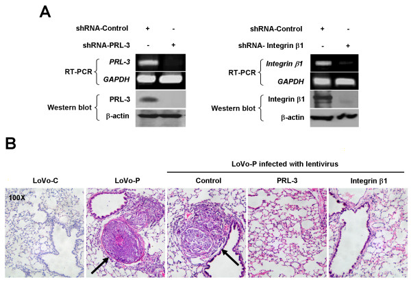
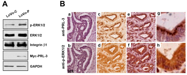
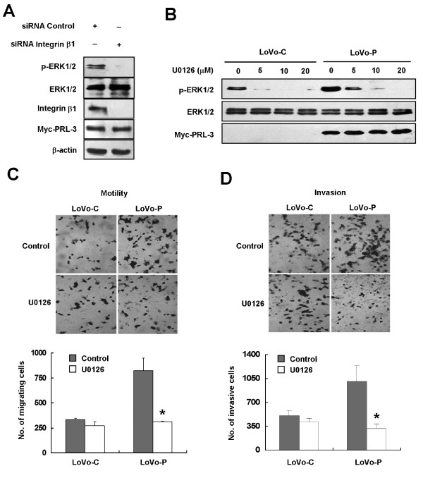
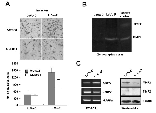
Results: We demonstrated that PRL-3 associated with integrin beta1 and its expression was positively correlated with ERK1/2 phosphorylation in colon cancer tissues. Depletion of integrin beta1 with siRNA, not only abrogated the activation of ERK1/2 stimulated by PRL-3, but also abolished PRL-3-induced motility and invasion of LoVo cells in vitro. Similarly, inhibition of ERK1/2 phosphorylation with U0126 or MMP activity with GM6001 also impaired PRL-3-induced invasion. In addition, PRL-3 promoted gelatinolytic activity of MMP2, and this stimulation correlated with decreased TIMP2 expression. Moreover, PRL-3-stimulated lung metastasis of LoVo cells in a nude mouse model was inhibited when integrin beta1 expression was interfered with shRNA.

Conclusion: Our results suggest that PRL-3's roles in motility, invasion, and metastasis in colon cancer are critically controlled by the integrin beta1-ERK1/2-MMP2 signaling.


Figures

Figure 1
Figure 1
PRL-3 interacts with integrin β1 and decreases its tyrosine phosphorylation in LoVo cells. (A) PRL-3 interacted with integrin β1. Equal amount of lysates (500 μg of protein) from LoVo-P cells, which stably expressed myc-tagged human PRL-3, and control LoVo-C cells were immunoprecipitated with anti-myc antibody, followed by Western blot with anti-integrin β1 antibody and anti-PRL-3 antibody. Expression of integrin β1 and PRL-3 in the lysates (50 μg of protein) was shown as Input. GAPDH protein expression was shown as a loading control. Molecular weight was shown. (B) PRL-3 was colocalized with integrin β1. LoVo cells were transiently transfected with GFP-PRL-3 (green). Twenty-four hours after transfection, cells were fixed, stained with an anti-integrin β1 antibody (red), and observed under a laser confocal microscope. The white arrow in Merge (4×) indicates the colocalization of PRL-3 with integrin β1 (yellow). (C) PRL-3 decreased tyrosine phosphorylated integrin β1. Equal amount of lysates (500 μg of preotein) from LoVo-C and LoVo-P cells were immunoprecipitated with anti-phosphotyrosine antibody or IgG control. The precipitates were subjected to Western blot with anti-integrin β1.
Figure 2
Figure 2
Integrin β1 mediates PRL-3-induced cell motility and invasion. (A) LoVo-P cells were treated with siRNA specific for integrin β1 or control. Seventy-two hours after transfection, cells were harvested. Equal amount of protein lysates (50 μg of protein) was analyzed for integrin β1 protein expression and RNA was extracted for RT-PCR. (B) and (C) Integrin β1 is required for PRL-3-induced cell motility and invasion. Cells were transfected with indicated siRNAs as in (A). Seventy-two hours after transfection, cells were analyzed for their motility and invasion with the use of transwell chambers. Cells were suspended in serum-free medium and loaded at a density of 2.5 × 10⁴ to an insert of a transwell chamber, and those migrating or invading to the underside of filtera were stained and counted after 24 h (B, motility assay) or 48 h (C, invasion assay). Top panel: Representative illustrations for motility and invasion assays (original magnification, × 200). Bottom panel: Quantification of migrating and invasive cells. Values were the total number of stained cells. The experiments were repeated at least three times independently. Error bars represent standard errors of the mean value (*, P < 0.05).
Figure 3
Figure 3
Depletion of integrin β1 or PRL-3 abrogates PRL-3-promoted metastasis in vivo. LoVo-P cells were infected with lentivirus interfering with PRL-3, integrin β1, or control respectively at a multiplicity of infection value of 100 for 48 h, or left untreated. (A) Validation of silencing efficiency of Lentivirus by RT-PCR and Western blot. (B) Each nude BALB/c mouse was injected via the tail vein with 2.5 × 10⁶ LoVo-C or LoVo-P cells. Two months later, mice were sacrificed, and 4-μm paraffin slices of liver and lung tissues were stained with hematoxylin and eosin dyes and examined under a light microscope. Arrows indicate the presence of a metastatic tumor in a lung slice.
Figure 4
Figure 4
Correlation between PRL-3 level and ERK phosphorylation. (A) PRL-3 enhanced ERK1/2 phosphorylation in LoVo cells. Equal numbers of LoVo-C and LovO-P cells were cultured for 24 h with complete medium. Cells were harvested and lyzed. Then cell lysates (50 μg of protein) were subjected to Western blot with antibodies against p-ERK1/2, ERK1/2, Myc-tag (for Myc-PRL-3), integrin β1, and GAPDH, respectively. (B) Expression of PRL-3 and p-ERK1/2 in primary lesions of human colon cancer tissues was analyzed by an immunohistochemical assay. Two consecutive 4-μm paraffin-embedded slices of the same tissue sample were probed with anti-PRL-3 (a, c, e, g) and anti-p-ERK1/2 (b, d, f, h), respectively. The representative negative staining of PRL-3 and p-ERK1/2 in the consecutive slices was shown in a and b, and the representative positive staining of PRL-3 and p-ERK1/2 was shown in the c, d, e, f, g, h (c and d, without HE counterstaining; e, f, g, h, with HE counterstaining; g and h, enlarged views of e and f, respectively).
Figure 5
Figure 5
Integrin β1 mediates PRL-3-induced ERK1/2 activation. (A) Knockdown of integrin β1 abolished PRL-3-induced ERK1/2 phosporylation. LoVo-P cells were treated with siRNA against integrin β1 or control for 72 h, and then lysates (50 μg of protein) were subjected to Western blot to analyze the expression of p-ERK1/2, ERK1/2, Myc-tag (for Myc-PRL-3), integrin β1, and β-actin (B) U0126 inhibited PRL-3-induced ERK1/2 phosporylation in a dose-dependent manner. Twenty-four hours after plating, LoVo-C and LoVo-P cells were treated with indicated concentration of U0126 for 1 h, and their lysates (50 μg of protein) were analyzed for p-ERK1/2, ERK1/2 and Myc-tag (for Myc-PRL-3) by Western blot. (C) and (D) Inhibition of ERK1/2 activity by U0126 abolished PRL-3-induced cell motility and invasion. LoVo-C and LoVo-P cells were treated with 10 μM of U0126 for 1 h, and then subjected to motility and invasion assays as described in Figure 2B and 2C, respectively. Values were the total number of stained cells. The experiments were repeated at least three times independently. Error bars represent standard errors of the mean value (*, P < 0.05).
Figure 6
Figure 6
PRL-3 promotes invasion by enhancing MMP2 activity. (A) MMP activity was required for PRL-3-mediated invasion. Cells were treated with 25 μM of GM6001 or DMSO (control) for 2 h, and analyzed for their invasion abilities with Matrigel-coated transwell chambers. The invasion assay was performed for three times independently. Top: Representative illustrations of LoVo cells invading to the underside of filters. Bottom: Total number of invasive cells. The experiments were repeated three times independently. Error bars represent standard errors of the mean value (*, P < 0.05). (B) PRL-3 increased gelatin hydrolytic activity of MMP2 in LoVo cells. Equal amount of conditioned serum-free media of LoVo-C and LoVo-P cells was subjected to a zymographic assay as described in Experimental Procedures. A small aliquot of calf serum containing MMP9 and MMP2 was included as the positive control. Bright bands contrasting to dark background represented the hydrolysis area of gelatin catalyzed by the MMP at the same molecular weight. (C) PRL-3 affected MMP2 and TIMP2 expression at both mRNA and protein levels. Equal amount of cell lysates (50 μg of protein) and RNA samples were subjected to Western blot and RT-PCR assays to analyze the expression of MMP2 and TIMP2, respectively.

Similar articles

Cited by

References

    1. Zeng Q, Hong W, Tan YH. Mouse PRL-2 and PRL-3, two potentially prenylated protein tyrosine phosphatases homologous to PRL-1. Biochem Biophys Res Commun. 1998;244:421–427. doi: 10.1006/bbrc.1998.8291. - DOI - PubMed
    1. Matter WF, Estridge T, Zhang C, Belagaje R, Stancato L, Dixon J, Johnson B, Bloem L, Pickard T, Donaghue M. Role of PRL-3, a human muscle-specific tyrosine phosphatase, in angiotensin-II signaling. Biochem Biophys Res Commun. 2001;283:1061–1068. doi: 10.1006/bbrc.2001.4881. - DOI - PubMed
    1. Fagerli UM, Holt RU, Holien T, Vaatsveen TK, Zhan F, Egeberg KW, Barlogie B, Waage A, Aarset H, Dai HY. Overexpression and involvement in migration by the metastasis-associated phosphatase PRL-3 in human myeloma cells. Blood. 2008;111:806–815. doi: 10.1182/blood-2007-07-101139. - DOI - PMC - PubMed
    1. Parker BS, Argani P, Cook BP, Liangfeng H, Chartrand SD, Zhang M, Saha S, Bardelli A, Jiang Y, St Martin TB. Alterations in vascular gene expression in invasive breast carcinoma. Cancer Res. 2004;64:7857–7866. doi: 10.1158/0008-5472.CAN-04-1976. - DOI - PubMed
    1. Rouleau C, Roy A, St Martin T, Dufault MR, Boutin P, Liu D, Zhang M, Puorro-Radzwill K, Rulli L, Reczek D. Protein tyrosine phosphatase PRL-3 in malignant cells and endothelial cells: expression and function. Mol Cancer Ther. 2006;5:219–229. doi: 10.1158/1535-7163.MCT-05-0289. - DOI - PubMed

Publication types

MeSH terms

Substances

LinkOut - more resources