. 2009 Nov 25:10:556.
doi: 10.1186/1471-2164-10-556.

Colorectal cancer cell-derived microvesicles are enriched in cell cycle-related mRNAs that promote proliferation of endothelial cells

Affiliations

Colorectal cancer cell-derived microvesicles are enriched in cell cycle-related mRNAs that promote proliferation of endothelial cells

Bok Sil Hong et al. BMC Genomics. .

Abstract

Background: Various cancer cells, including those of colorectal cancer (CRC), release microvesicles (exosomes) into surrounding tissues and peripheral circulation. These microvesicles can mediate communication between cells and affect various tumor-related processes in their target cells.

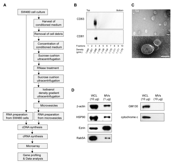
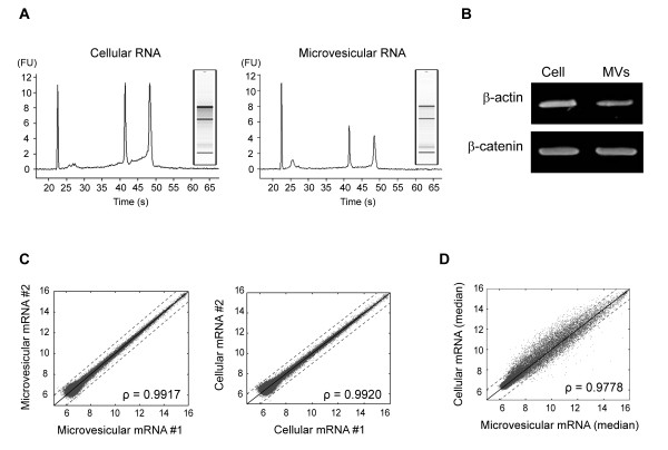
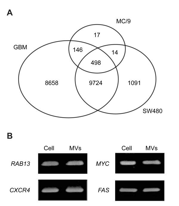
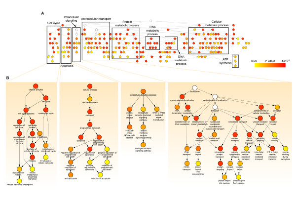
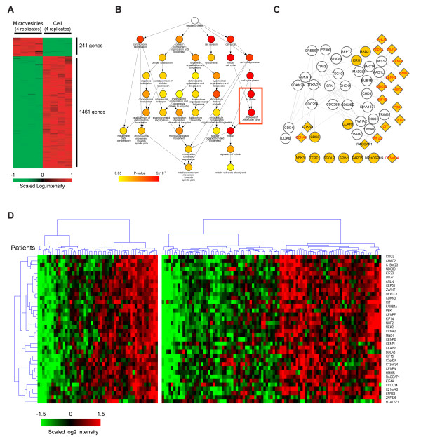
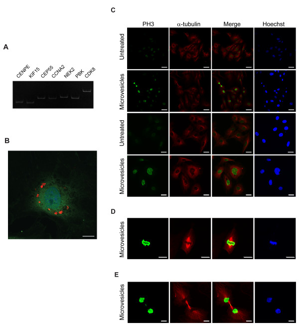
Results: We present potential roles of CRC cell-derived microvesicles in tumor progression via a global comparative microvesicular and cellular transcriptomic analysis of human SW480 CRC cells. We first identified 11,327 microvesicular mRNAs involved in tumorigenesis-related processes that reflect the physiology of donor CRC cells. We then found 241 mRNAs enriched in the microvesicles above donor cell levels, of which 27 were involved in cell cycle-related processes. Network analysis revealed that most of the cell cycle-related microvesicle-enriched mRNAs were associated with M-phase activities. The integration of two mRNA datasets showed that these M-phase-related mRNAs were differentially regulated across CRC patients, suggesting their potential roles in tumor progression. Finally, we experimentally verified the network-driven hypothesis by showing a significant increase in proliferation of endothelial cells treated with the microvesicles.

Conclusion: Our study demonstrates that CRC cell-derived microvesicles are enriched in cell cycle-related mRNAs that promote proliferation of endothelial cells, suggesting that microvesicles of cancer cells can be involved in tumor growth and metastasis by facilitating angiogenesis-related processes. This information will help elucidate the pathophysiological functions of tumor-derived microvesicles, and aid in the development of cancer diagnostics, including colorectal cancer.


Figures

Figure 1
Figure 1
Purification and characterization of microvesicles derived from SW480 cells. A) Overview of the procedure used to identify microvesicular and cellular mRNA from SW480 cells. B) Western blot of microvesicular marker proteins CD63 and CD81 in fractions from iodixanol density gradients. C) Electron microscopy of the purified microvesicles. Bars represent 100 nm. D) Western blot of β-actin, HSP90, Ezrin, and Rab5A (microvesicular proteins), and GM130 (a protein found in the cis-Golgi apparatus), and cytochrome c (a mitochondrial protein found in apoptotic bodies). Neither GM130 nor cytochrome c were detected in microvesicles, despite using 10 times more microvesicles (10 μg) than the amount used to detect other microvesicular proteins (1 μg). The symbols of WCL and MVs represent whole cell lysate and microvesicles, respectively.
Figure 2
Figure 2
Isolation and transcriptomic analysis of microvesicular RNA. A) Bioanalyzer results of total RNA isolated from microvesicles and SW480 cells. The third and fourth peaks correspond to 18S and 28S rRNA, respectively. Images of gel electrophoresis are also shown. B) Gel electrophoresis of microvesicular and cellular β-actin and β-catenin mRNA amplified by RT-PCR. C) Scatterplots of microvesicular and cellular mRNA expression levels showing log2 intensities in the first and the second arrays (Pearson's correlation coefficients: ρ = 0.9917 and 0.9920 for microvesicular RNA and cellular RNA, respectively). D) Scatterplot of median log2 intensities of the four microvesicle replicates versus those of their donor cells (ρ = 0.9778).
Figure 3
Figure 3
Comparison with other microarray studies and validation of the microvesicular and cellular mRNAs detected in our study. A) Comparison among the number of mRNAs detected in microvesicles derived from mouse mast cells (MC/9) [14], glioblastoma (GBM) [15] and human SW480 CRC cells. B) Gel electrophoresis of amplified microvesicular and cellular mRNA. Presence of several microvesicular mRNAs including RAB13, CXCR4, MYC, and FAS were confirmed using RT-PCR. The symbol of MVs represents microvesicles.
Figure 4
Figure 4
Cellular processes overrepresented by the mRNAs detected in microvesicles. A) A GO tree obtained from BiNGO showing the hierarchy of GOBPs overrepresented by mRNAs present in microvesicles (P < 0.05; see the color bar). Node colors represent the statistical significance of functional enrichment of the corresponding GOBPs. The white nodes (P > 0.05) were added to show the relationships among their downstream nodes. The overrepresented GOBPs were categorized into several groups of cellular processes, each of which is indicated by a box. B) The four GOBP groups (cell cycle, apoptosis, intracellular signaling, and intracellular transport) related to tumor progression are shown in detail.
Figure 5
Figure 5
A biological network describing the cellular processes overrepresented by microvesicle-enriched mRNAs. A) A heat map illustrating the expression patterns of the 1,702 genes that exhibited a more than 2-fold difference in the mRNA level between microvesicles and cells. For clarity, the expression of each is scaled to a mean of zero with unit variance across all eight replicates. The colors represent the relative increase (red) or decrease (green) in the expression level of the corresponding gene in microvesicles compared with the level in cells: 241 and 1,461 genes were overexpressed in microvesicles and in cells, respectively. B) A GO subtree showing cell cycle-related GOBPs overrepresented by the 241 microvesicle-enriched genes. The color scheme of Figure 4 is repeated. Notably, cell cycle-related GOBPs were strongly overrepresented by 27 mRNAs, as indicated by the dark-red node colors (see text for details). C) A hypothetical network reconstructed using the 27 microvesicle-enriched mRNAs belonging to the cell cycle-related GOBPs in (B) and their first and second interaction neighbors obtained from protein-protein interaction databases (see Materials and methods). The yellow nodes represent the 27 cell cycle-related microvesicle-enriched genes: 20 of the 27 genes closely interact with cell cycle-related processes. The lines between the nodes represent protein-protein interactions. The network shows that the microvesicle-enriched genes closely interact with key pathways in the cell cycle. D) Cluster heat map comprising 36 of the 241 microvesicle-enriched mRNAs showing consistent differential expression patterns across patients in two independent CRC datasets (GSE2109 and GSE5206). The diamond nodes in the network (C) represent the 17 nodes shared by the 27 cell cycle-associated mRNAs and 36 mRNAs; of the 17 nodes, 15 (labeled in red) are associated with M-phase-related processes.
Figure 6
Figure 6
Stimulation of endothelial cell proliferation by microvesicles. A) Gel electrophoresis of amplified microvesicular M-phase-related transcripts. Presence of CENPE, KIF15, CEP55, CCNA2, NEK2, PBK and CDK8 were confirmed using RT-PCR. B) Immunofluorescence of HUVECs treated with microvesicles labeled with PKH26 (red). Cells were stained with anti-calnexin antibody, an endoplasmic reticulum marker protein (green). C-E) Immunostaining of HUVECs with anti-phospho-histone H3 (green) and anti-α-tubulin antibodies (red). After 12 hours, microvesicle-treated endothelial cells were at metaphase (D) and cytokinesis with mitotic spindle formation (E). None of the untreated control endothelial cells were in metaphase or cytokinesis stages. Scale bars represent 10 μm (B, D, E) and 40 μm (C). The symbol of PH3 represents phospho-histone H3.

References

    1. Fevrier B, Raposo G. Exosomes: endosomal-derived vesicles shipping extracellular messages. Curr Opin Cell Biol. 2004;16(4):415–421. doi: 10.1016/j.ceb.2004.06.003. - DOI - PubMed
    1. Kim CW, Lee HM, Lee TH, Kang C, Kleinman HK, Gho YS. Extracellular membrane vesicles from tumor cells promote angiogenesis via sphingomyelin. Cancer Res. 2002;62(21):6312–6317. - PubMed
    1. Théry C, Zitvogel L, Amigorena S. Exosomes: composition, biogenesis and function. Nat Rev Immunol. 2002;2(8):569–79. - PubMed
    1. Ratajczak J, Wysoczynski M, Hayek F, Janowska-Wieczorek A, Ratajczak MZ. Membrane-derived microvesicles: important and underappreciated mediators of cell-to-cell communication. Leukemia. 2006;20(9):1487–1495. doi: 10.1038/sj.leu.2404296. - DOI - PubMed
    1. Choi DS, Lee JM, Park GW, Lim HW, Bang JY, Kim YK, Kwon KH, Kwon HJ, Kim KP, Gho YS. Proteomic analysis of microvesicles derived from human colorectal cancer cells. J Proteome Res. 2007;6(12):4646–4655. doi: 10.1021/pr070192y. - DOI - PubMed

Publication types