. 2010 Jun;223(2):422-31.
doi: 10.1016/j.expneurol.2009.11.005. Epub 2009 Nov 18.

Type 1 diabetes exaggerates features of Alzheimer's disease in APP transgenic mice

Affiliations

Type 1 diabetes exaggerates features of Alzheimer's disease in APP transgenic mice

Corinne G Jolivalt et al. Exp Neurol. 2010 Jun.

Abstract

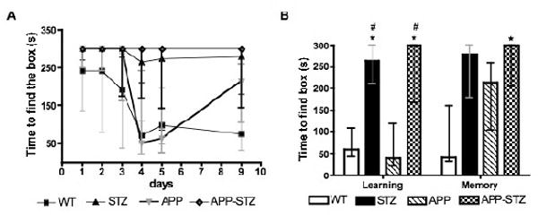
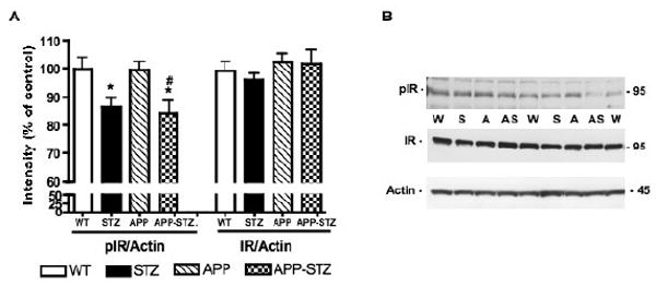
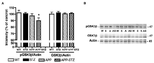
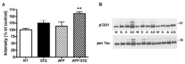
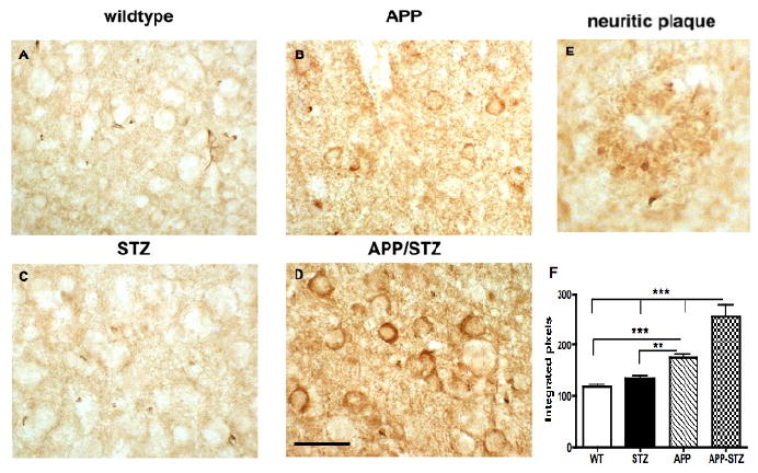
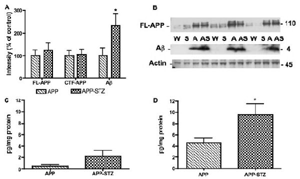
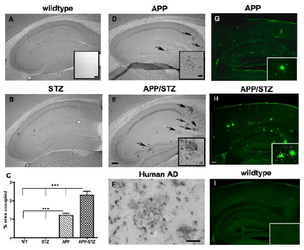
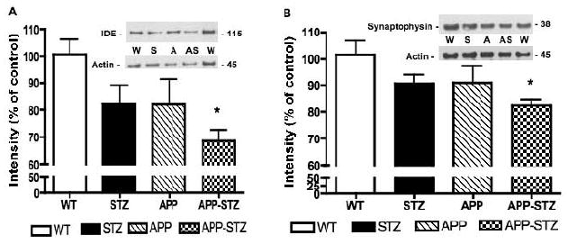
A number of studies suggest an association between Alzheimer's disease (AD) and diabetes: AD patients show impaired insulin function, whereas cognitive deficits and increased risk of developing AD occur in diabetic patients. The reasons for the increased risk are not known. Recent studies of disturbances in the insulin-signaling pathway have revealed new perspectives on the links between AD and Type 1 diabetes with a particular focus on glycogen synthase-kinase-3 (GSK3). We have therefore characterized a mouse model of combined insulin-deficient diabetes and AD and find that diabetes exaggerated defects in the brain of APP transgenic mice. Mice with combined APP overexpression and diabetes showed a decreased insulin receptor activity and an increased GSK3beta activity. Concomitantly, tau phosphorylation and number of Abeta plaques, the two pathologic hallmarks of AD, were increased in the brain of diabetic-APP transgenic mice. Our results indicate that the pathologic features of AD are exaggerated in the brain of APP transgenic mice that have concurrent insulin-deficient diabetes, and underscore a possible mechanism of brain dysfunction common to AD and diabetes.


Figures

Figure 1
Figure 1. Barnes maze task
A: Time course of the learning and memory assessment using the Barnes maze test. B: Time to find the escape box at day 4 and 9: Learning was assessed on day 4 after 4 consecutive testing sessions and memory was assessed on day 9 after 3 days without testing. Data are represented as median ± interquartiles. * p< 0.05 and # p<0.05 by Kruskal-Wallis for nonparametric data followed by Dunn's post hoc test vs WT and APP group, respectively. n=8 for WT and STZ groups, n=5 for APP and APP-STZ groups.
Figure 2
Figure 2. Phosphorylation level of insulin receptor in mouse brain
A: Intensity of bands corresponding to phosphorylated insulin receptor (pIR) and insulin receptor (IR) normalized to the intensity of bands corresponding to actin. Data are represented as mean+sem. *p<0.05 vs WT, #p<0.05 vs APP by one-way ANOVA followed by Bonferroni's post hoc test. n=8 for WT and STZ groups, n=5 for APP group and n=4 for APP-STZ group. B: Western blots of brain homogenates from Wild type (W), STZ-diabetic (S), APP (A) and APP-STZ (AS) mice for phosphorylated insulin receptor (pIR), total insulin receptor (IR) and actin.
Figure 3
Figure 3. Phosphorylation level of GSK3β in mouse brain
A: Intensity of bands corresponding to phosphorylated GSK3β (pGSK3β) and GSK3β normalized to the intensity of bands corresponding to actin. Data are represented as mean+sem. *p<0.05 vs WT by one-way ANOVA followed by Bonferroni's post hoc test. n=8 for WT and STZ groups, n=5 for APP group and n=4 for APP-STZ group B: Western blots of brain homogenates from Wild type (W), STZ-diabetic (S), APP (A) and APP-STZ (AS) mice for phosphorylated GSK3β (pGSK3β), total GSK3β (GSK3β) and actin.
Figure 4
Figure 4. Phosphorylated tau in mouse brain
A: Intensity of bands corresponding to phosphorylated tau at threonine 231 (p T231) normalized to the intensity of bands corresponding to total tau (pan Tau). Data are represented as mean+sem. **p<0.01 vs WT by one-way ANOVA followed byBonferroni's post hoc test. n=8 for WT and STZ groups, n=5 for APP group and n=4 for APP-STZ group B: Western blots of brain homogenates from Wild type (W), STZ-diabetic (S), APP (A) and APP-STZ (AS) mice for phosphorylated tau at threonine 231 (p T231) and total tau (pan Tau).
Figure 5
Figure 5. Phosphorylated tau in mouse brain
A-D: Phosphorylated tau (PHF1) immunoreactivity in mouse hippocampus illustrating somatodendritic accumulation of phosphorylated tau in APP-STZ mice brain (D), bar=40 μm. E: Phosphorylated tau immunoreactivity associated with neuritic plaque in APP-STZ mice hippocampus. F: Quantitative analysis of phosphorylated tau immunoreactivity in brain from Wild type (WT), STZ-diabetic (STZ), APP and APP-STZ mice. **p<0.01, ***p<0.01 by one-way ANOVA followed by Bonferroni's post hoc test.
Figure 6
Figure 6. Amyloid β level in mouse brain
A: Intensity of bands corresponding to full length APP (FL-APP), C terminal fragment-APP (CTF-APP) and amyloid β (Aβ) normalized to the intensity of bands corresponding to actin. Data are represented as mean+sem. *p<0.05 vs APP by unpaired t test. n=5 for APP group and n=4 for APP-STZ group. B: Western blots of brain homogenates from Wild type (W), STZ-diabetic (S), APP (A) and APP-STZ (AS) mice for full length APP (FL-APP), amyloid β (Aβ) and actin. C: Aβ1-40 levels in brain homogenates from APP and APP-STZ mice using an Elisa assay for human amyloid β1–40. Data are represented as mean+sem. D:1-42 levels in brain homogenates from APP and APP-STZ mice using an Elisa assay for human amyloid β1–42. Data are represented as mean+sem. *p<0.05 vs APP by unpaired t test. n=5 for APP group and n=4 for APP-STZ group.
Figure 7
Figure 7. Amyloid β-immunoreactive plaques in mice hippocampus
Amyloid β-immunoreactive plaques in hippocampus from Wild type (A), STZ-diabetic (STZ, B), APP (D) and diabetic-APP (APP/STZ, E) mice after 12 weeks of diabetes. Arrows point at amyloid β-immunoreactive plaques, bar=200μm. Inset in A: Specificity of immunostaining by omission of the primary antibody, bar=200 μm. Insets in D and E: magnification of amyloid β-immunoreactive plaques in APP and APP-STZ mice hippocampus, respectively, bar=40μm. C: Quantification of amyloid β-immunoreactive plaques as percent area occupied by plaques in mouse hippocampus. Data are represented as mean+sem. ***p<0.001 by one-way ANOVA followed by Bonferroni's post hoc test. n=8 for WT and STZ groups, n=5 for APP group and n=4 for APP-STZ group. F: Amyloid β-immunoreactive plaques in the brain from a patient diagnosed with late stage AD, bar=40μm. G-H: Mature plaques were intensively labeled with Thioflavine-S in hippocampus and cortex from APP and APP/STZ mice, respectively but not in Wildtype (I), bar=200 μm. Insets in G, H and I: Magnification of hippocampus containing plaques for APP (G) and APP/STZ (H) mice and no plaques for WT mouse (I).
Figure 8
Figure 8. IDE and synaptophysin protein level in mouse brain
A: Intensity of bands corresponding to IDE normalized to the intensity of bands corresponding to actin. Data are represented as mean+sem. *p<0.05 vs WT by one-way ANOVA followed by Bonferroni's post hoc test. Western blots of brain homogenates from Wild type (W), STZ-diabetic (S), APP (A) and APP-STZ (AS) mice for IDE and actin. B: Intensity of bands corresponding to synaptophysin normalized to the intensity of bands corresponding to actin. Data are represented as mean+sem. *p<0.05 vs WT by one-way ANOVA followed by Bonferroni's post hoc test. Western blots of brain homogenates from Wild type (W), STZ-diabetic (S), APP (A) and APP-STZ (AS) mice for synaptophysin and actin. n=8 for WT and STZ groups, n=5 for APP group and n=4 for APP-STZ group.

References

    1. Akomolafe A, Beiser A, Meigs JB, Au R, Green RC, Farrer LA, Wolf PA, Seshadri S. Diabetes mellitus and risk of developing Alzheimer disease: results from the Framingham Study. Arch Neurol. 2006;63:1551–1555. - PubMed
    1. Barnes CA. Memory deficits associated with senescence: a neurophysiological and behavioral study in the rat. J Comp Physiol Psychol. 1979;93:74–104. - PubMed
    1. Biessels GJ, Deary IJ, Ryan CM. Cognition and diabetes: a lifespan perspective. Lancet Neurol. 2008;7:184–190. - PubMed
    1. Braak H, Braak E. Neuropathological stageing of Alzheimer-related changes. Acta Neuropathol. 1991;82:239–259. - PubMed
    1. Brownlees J, Irving NG, Brion JP, Gibb BJ, Wagner U, Woodgett J, Miller CC. Tau phosphorylation in transgenic mice expressing glycogen synthase kinase-3beta transgenes. Neuroreport. 1997;8:3251–3255. - PubMed

Publication types

MeSH terms