Review
. 2010 Feb;37(2):259-66.
doi: 10.1016/j.nbd.2009.11.009. Epub 2009 Nov 26.

Promoting axonal rewiring to improve outcome after stroke

Affiliations
Review

Promoting axonal rewiring to improve outcome after stroke

Larry I Benowitz et al. Neurobiol Dis. 2010 Feb.

Abstract

A limited amount of functional recovery commonly occurs in the weeks and months after stroke, and a number of studies show that such recovery is associated with changes in the brain's functional organization. Measures that augment this reorganization in a safe and effective way may therefore help improve outcome in stroke patients. Here we review some of the evidence for functional and anatomical reorganization under normal physiological conditions, along with strategies that augment these processes and improve outcome after brain injury in animal models. These strategies include counteracting inhibitors of axon growth associated with myelin, activating neurons' intrinsic growth state, enhancing physiological activity, and having behavioral therapy. These approaches represent a marked departure from the recent focus on neuroprotection and may provide a more effective way to improve outcome after stroke.


Figures
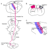
Figure 1
Figure 1. Patterns of axonal sprouting after stroke
(A) Schematic view of motor cortex (red) projections from the brain to the brainstem and cervical spinal cord. In each panel the normal axonal projections from motor cortex are in solid lines. The projections from the cortical region lost from the stroke are in gray dashed lines. The projections that form after stroke are in red dashed lines. In (A), a stroke induces axonal sprouting from contralateral motor cortex into the red nucleus (Papadopoulos et al., 2002) or into the ipsilateral cervical spinal cord (Chen et al., 2002; Zai et al., 2009). These projections form in areas that were denervated by the loss of the stroked cortex. In (B), a stroke induces axonal sprouting from contralateral motor cortex into the ipsilateral striatum and contralateral peri-infarct cortex (Carmichael and Chesselet, 2002). These are areas in which the projections from the stroke site are lost. Stroke also induces several patterns of axonal sprouting in cortex adjacent or ipsilateral to the stroke site, shown in (C). New projections from retrosplenial cortex to peri-infarct cortex form after small strokes in motor cortex (Brown et al., 2009). Somatosensory cortex establishes new direct projections to the ventral premotor cortex. This pattern of axonal sprouting, identified in the squirrel monkey, establishes a long-distance projection from parietal to frontal lobe (Dancause et al., 2005). Premotor cortex in the rate is a site of motor remapping that correlates with functional recovery after cortical injury (Conner et al., '05). Stroke also causes a substantial axonal sprouting response in peri-infarct cortex near the stroke site (Carmichael et al., 2001).
Figure 2
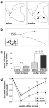
Figure 2. Inosine enhances axonal rewiring and augments functional recovery in an animal model of stroke
a Camera lucida drawings of BDA-labeled corticospinal tract (CST) fibers that originate in the uninjured hemisphere and project to the side of the spinal cord denervated by injury to the contralateral sensorimoter cortex. Rats were treated with either saline- (left) or inosine (right). b Higher-magnification camera lucida tracing of a fiber in a. Inset shows the presence of synaptic bouton-like structures. c Quantitation of ipsilaterally projecting CST fibers ≥ 40 μm in length in the transverse plane in the denervated dorsal funiculus and gray matter, respectively. Results are normalized by the intensity of staining in the intact CST and are reported as the number of labeled axons per mm length of spinal cord. Inosine does not promote CST axon growth in the absence of brain injury, but amplifies the amount of axon sprouting after a stroke. d Inosine enhances functional recovery. Animals were trained to retrieve food pellets through a restricted opening with either paw prior to surgery and were then tested beginning one week later by a blinded observer. Scores are reported as percentage of pre-operative performance. *, **, ***: Differences significant at P < 0.05, P < 0.01, P < 0.001, respectively. Error bars represent SEM. (from Zai et al., 2009).

Similar articles

Cited by

References

    1. Atwal JK, et al. PirB is a functional receptor for myelin inhibitors of axonal regeneration. Science. 2008;322:967–70. - PubMed
    1. Benowitz LI, et al. Axon outgrowth is regulated by an intracellular purine-sensitive mechanism in retinal ganglion cells. J Biol Chem. 1998;273:29626–34. - PubMed
    1. Benowitz LI, et al. The pattern of GAP-43 immunostaining changes in the rat hippocampal formation during reactive synaptogenesis. Brain Research Molecular Brain Research. 1990;8:17–23. - PubMed
    1. Benowitz LI, Routtenberg A. GAP-43: an intrinsic determinant of neuronal development and plasticity. Trends in Neurosciences. 1997;20:84–91. - PubMed
    1. Benson MD, et al. Ephrin-B3 is a myelin-based inhibitor of neurite outgrowth. Proc Natl Acad Sci U S A. 2005;102:10694–9. - PMC - PubMed

Publication types

MeSH terms