. 2010 Mar;93(3):303-11.
doi: 10.1016/j.nlm.2009.11.004. Epub 2009 Nov 29.

Unimpaired trace classical eyeblink conditioning in Purkinje cell degeneration (pcd) mutant mice

Affiliations

Unimpaired trace classical eyeblink conditioning in Purkinje cell degeneration (pcd) mutant mice

Kevin L Brown et al. Neurobiol Learn Mem. 2010 Mar.

Abstract

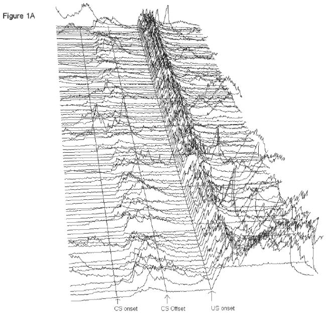
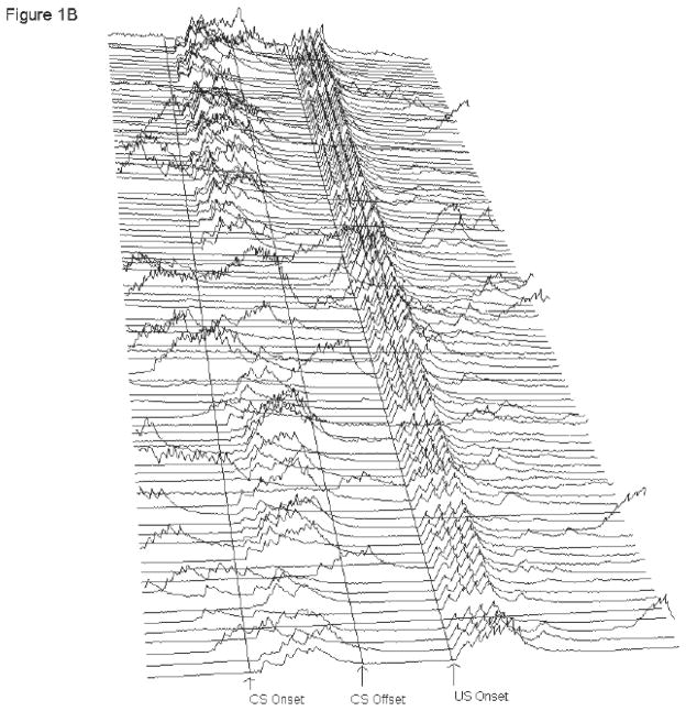
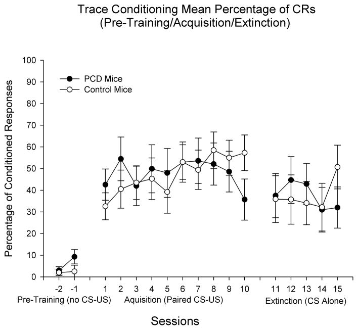
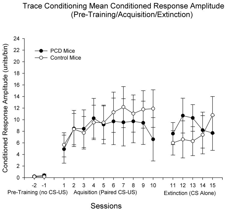
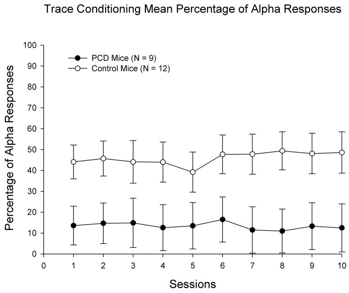
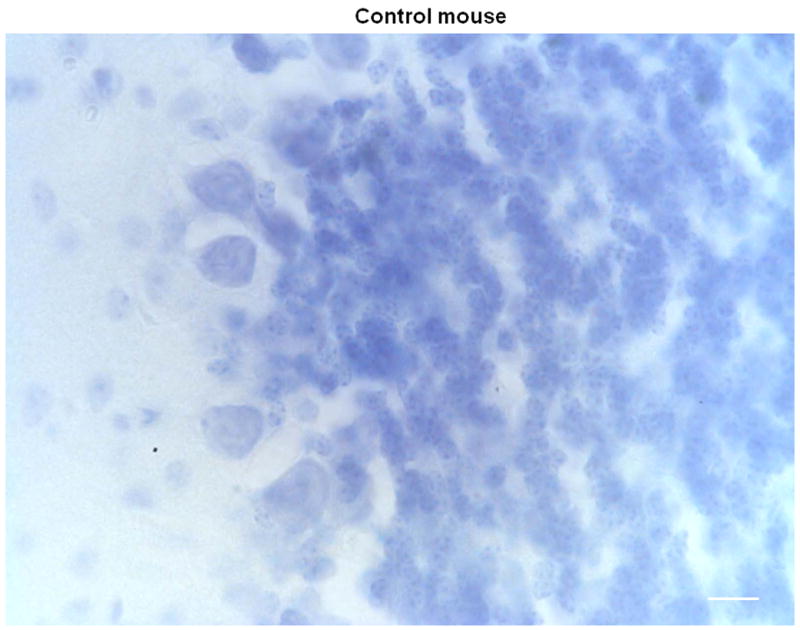
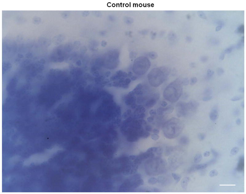
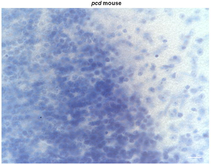
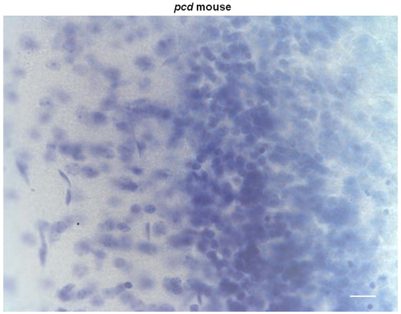
Young adult Purkinje cell degeneration (pcd) mutant mice, with complete loss of cerebellar cortical Purkinje cells, are impaired in delay eyeblink classical conditioning. In the delay paradigm, the conditioned stimulus (CS) overlaps and coterminates with the unconditioned stimulus (US), and the cerebellar cortex supports normal acquisition. The ability of pcd mutant mice to acquire trace eyeblink conditioning in which the CS and US do not overlap has not been explored. Recent evidence suggests that cerebellar cortex may not be necessary for trace eyeblink classical conditioning. Using a 500 ms trace paradigm for which forebrain structures are essential in mice, we assessed the performance of homozygous male pcd mutant mice and their littermates in acquisition and extinction. In contrast to results with delay conditioning, acquisition of trace conditioning was unimpaired in pcd mutant mice. Extinction to the CS alone did not differ between pcd and littermate control mice, and timing of the conditioned response was not altered by the absence of Purkinje cells during acquisition or extinction. The ability of pcd mutant mice to acquire and extinguish trace eyeblink conditioning at levels comparable to controls suggests that the cerebellar cortex is not a critical component of the neural circuitry underlying trace conditioning. Results indicate that the essential neural circuitry for trace eyeblink conditioning involves connectivity that bypasses cerebellar cortex.


Figures

Figure 1
Figure 1
A and B: Electromyography (EMG) recorded from eye muscles (orbicularis oculi) of the left upper eyelid during trace eyeblink conditioning. Each line represents EMG activity from an individual trial (1–100, with Trial 1 represented at the bottom of each figure and Trial 100 represented at the top of each figure) from Session 6 of 500-ms trace eyeblink classical conditioning. Total trial length was 1,350 ms. Lines are drawn to approximate the onset of the conditioned stimulus (CS) and unconditioned stimulus (US). There were 249 ms in the pre-CS period before CS onset. CS onset is marked, and then there were 500 ms between CS onset and US onset (marked). There were 601 ms in the post-US period. A response was scored if it exceeded mean pre-CS activity by five standard deviations. Performance is shown for a pcd mouse (A) and a wildtype littermate (B) that were representative of their respective groups. For the pcd mouse shown here (A), 90 of the 100 trials were usable for analysis; the remaining 10 trials were excluded due to excessive pre-CS EMG activity. During paired CS-US trials this subject (A) performed 48% conditioned responses (CRs). Short-latency alpha (or ‘startle’) responses (0–60 ms after CS onset) occurred in 3% of the paired CS-US trials. For the wildtype littermate (B), 87 of the 100 trials were usable for analysis. During paired CS-US trials there were 68% CRs. Consistent with the higher percentage of short-latency alpha responses emitted in wildtypes relative to pcds, alpha responses occurred during 54% of the paired CS-US trials.
Figure 1
Figure 1
A and B: Electromyography (EMG) recorded from eye muscles (orbicularis oculi) of the left upper eyelid during trace eyeblink conditioning. Each line represents EMG activity from an individual trial (1–100, with Trial 1 represented at the bottom of each figure and Trial 100 represented at the top of each figure) from Session 6 of 500-ms trace eyeblink classical conditioning. Total trial length was 1,350 ms. Lines are drawn to approximate the onset of the conditioned stimulus (CS) and unconditioned stimulus (US). There were 249 ms in the pre-CS period before CS onset. CS onset is marked, and then there were 500 ms between CS onset and US onset (marked). There were 601 ms in the post-US period. A response was scored if it exceeded mean pre-CS activity by five standard deviations. Performance is shown for a pcd mouse (A) and a wildtype littermate (B) that were representative of their respective groups. For the pcd mouse shown here (A), 90 of the 100 trials were usable for analysis; the remaining 10 trials were excluded due to excessive pre-CS EMG activity. During paired CS-US trials this subject (A) performed 48% conditioned responses (CRs). Short-latency alpha (or ‘startle’) responses (0–60 ms after CS onset) occurred in 3% of the paired CS-US trials. For the wildtype littermate (B), 87 of the 100 trials were usable for analysis. During paired CS-US trials there were 68% CRs. Consistent with the higher percentage of short-latency alpha responses emitted in wildtypes relative to pcds, alpha responses occurred during 54% of the paired CS-US trials.
Figure 2
Figure 2
Trace eyeblink classical conditioning in Purkinje cell degeneration (pcd) mutant mice and wildtype littermate control mice. Percentage of conditioned responses (CRs) are displayed across the three phases of training: (1) pre-training (2 sessions) in which the animals were placed into the experimental apparatus without presentations of the CS or unconditioned stimulus (US); (2) acquisition training (10 daily 100-trial sessions) in which the CS preceded and reliably predicted the US; and (3) extinction training (5 daily 100-trial sessions) in which the CS was presented as in acquisition training, but without US presentations. The duration of pre-training sessions was equal to that of acquisition and extinction sessions (approximately 1 hour). CR incidence at all three phases of training does not differ as a function of group. As expected, modest declines in CR incidence are evident during CS-alone extinction training. Data in panels represent means plus and minus the standard error of the mean. The Ns differ in each phase: Phase 1 (pre-training) – N = 3 pcd; 4 control; Phase 2 (acquisition training) – N = 9 pcd; 12 control; Phase 3 (extinction training) – N = 9 pcd; 8 control.
Figure 3
Figure 3
Trace eyeblink classical conditioning in Purkinje cell degeneration (pcd) mutant mice and wildtype littermate control mice. Peak amplitude of conditioned responses (CRs) are displayed across the three phases of training: (1) pre-training (2 sessions) in which the animals were placed into the experimental apparatus without presentations of the CS or unconditioned stimulus (US); (2) acquisition training (10 daily 100-trial sessions) in which the CS preceded and reliably predicted the US; and (3) extinction training (5 daily 100-trial sessions) in which the CS was presented as in acquisition training, but without US presentations. The duration of pre-training sessions was equal to that of acquisition and extinction sessions (approximately 1 hour). As in the CR percentage measure, CR amplitude does not differ as a function of group in any of the three phases of training. Unlike controls, CR amplitude levels during extinction did not return to initial acquisition levels in pcd mice. Data represent means plus and minus the standard error of the mean. The Ns differ in each phase: Phase 1 (pre-training) – N = 3 pcd; 4 control; Phase 2 (acquisition training) – N = 9 pcd; 12 control; Phase 3 (extinction training) – N = 9 pcd; 8 control.
Figure 4
Figure 4
Percentage of alpha responding (responses occurring within the first 60 ms of CS onset) during acquisition training in Purkinje cell degeneration (pcd) mutant mice and wildtype littermate control mice. Robust group differences are evident, as the percentage of alpha responding in controls is substantially higher than in pcd mutant mice. Data represent means plus and minus the standard error of the mean. The Ns for each group are the same as reported in acquisition phases for the CR percentage and CR peak amplitude measures.
Figure 5
Figure 5
Representative coronal sections of the cerebellar cortex stained with Thionin in 2 control (A, B) and 2 pcd mice (C, D) magnified at 60x. Note the absence of Purkinje cells in the pcd sections (C, D), while Purkinje cells are abundant in the control sections (A, B). Scale bars = 20 μm.
Figure 5
Figure 5
Representative coronal sections of the cerebellar cortex stained with Thionin in 2 control (A, B) and 2 pcd mice (C, D) magnified at 60x. Note the absence of Purkinje cells in the pcd sections (C, D), while Purkinje cells are abundant in the control sections (A, B). Scale bars = 20 μm.

Similar articles

Cited by

References

    1. Aiba A, Kano M, Chen C, Stanton ME, Fox GD, Herrup K, Zwingman TA, Tonegawa S. Deficient cerebellar long-term depression and impaired motor learning in mGluR1 mutant mice. Cell. 1994;79:377–388. - PubMed
    1. Bao S, Chen L, Thompson RF. Classical eyeblink conditioning in two strains of mice: conditioned responses, sensitization, and spontaneous eyeblinks. Behavioral Neuroscience. 1998a;112:714–718. - PubMed
    1. Bao S, Chen L, Qiao X, Knusel B, Thompson RF. Impaired eye-blink conditioning in waggler, a mutant mouse with cerebellar BDNF deficiency. Learning and Memory. 1998b;5:355–364. - PMC - PubMed
    1. Buchanan SL, Thompson RH, Maxwell BL, Powell DA. Efferent connections of the medial prefrontal cortex in the rabbit. Experimental Brain Research. 1994;100:469–483. - PubMed
    1. Chen L, Bao S, Lockard JM, Kim JJ, Thompson RF. Impaired classical eyeblink conditioning in cerebellar-lesioned and Purkinje cell degeneration (pcd) mutant mice. The Journal of Neuroscience. 1996;16:2829–2838. - PMC - PubMed

Publication types